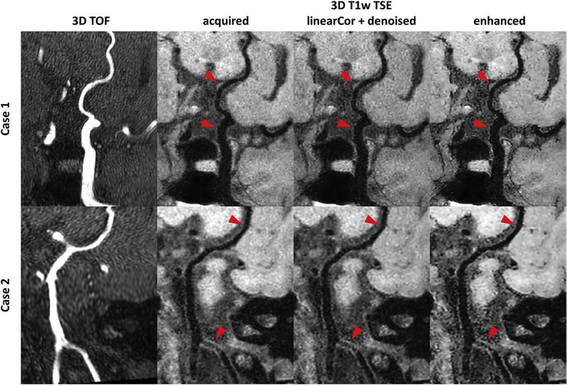
Figure 6:
Example curved planar reformatted (CPR) images from intracranial vessel scans on patients. The top case shows CPR images covering from the left internal carotid artery to posterior communicating artery, and the bottom case shows CPR images covering from vertebral arteries to right posterior cerebral artery. Different columns from left to right correspond to 3D time-of-flight (TOF) CPR images, the acquired CPR images with the optimized 3D T1 weighted (T1w) turbo spin echo (TSE) sequence, the linear enhanced CPR images with wavelet denoising, and the convolutional neural network (CNN) enhanced CPR images. The local vessel wall regions (red arrows) can be better delineated on the CNN enhanced images.

