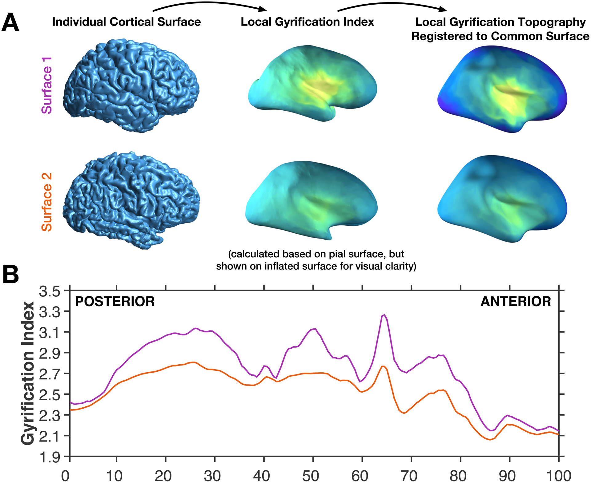
Figure 2. Illustration of the calculation of the anterior-posterior gradient.

(A) Individual brain pial surfaces are used to generate local gyrification index topography (based on Schaer et al., 2012), these are then resampled to the common space, through registration of the individual pial surface to the FreeSurfer standard surface. (B) The local gyrification index is then averaged across vertices for 200 coronal sections, shown for the same two example surfaces as in panel A.
