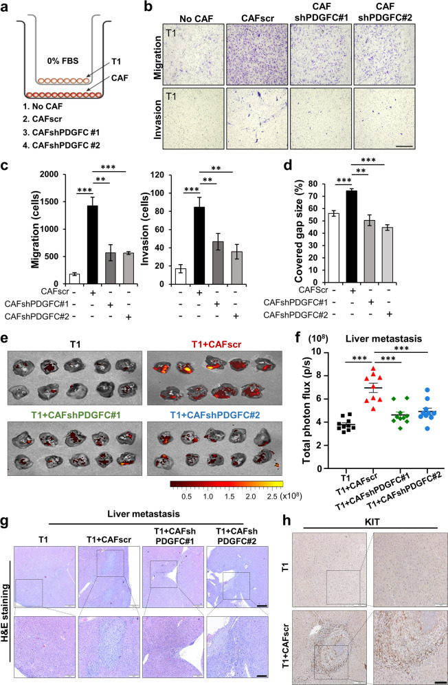
Fig. 4. PDGFC secreted from CAFs is required for GIST migration and invasion.
a–c Effects of stable PDGFC knockdown of CAFs on Transwell migration and invasion assays of T1 cells. Experimental design for the Transwell assays (a) and representative images of the migrated cells (b). The quantitative data (c) were generated with migrated cells that were counted by ImageJ software. p values were represented by ANOVA analysis. **p < 0.01, ***p < 0.001. d Wound healing assay in the indicated conditions. Data represents average of % covered gap size. p values were represented by ANOVA analysis. **p < 0.01, ***p < 0.001. e, f Spleen-to-liver metastasis model showing the effect of PDGFC secreted from CAFs. The mice were injected with mCherry-labeled T1, T1 + CAFscr, T1 + CAFshPDGFC #1, and T1 + CAFshPDGFC #2. IVIS images of metastatic liver (e) and quantification (f). Liver metastasis burden was quantified by total photon flux (p/s) on day 21 after the cell injection. p values were represented by ANOVA analysis. ***p < 0.001. Representative hematoxylin and eosin (H&E) images (g) and IHC images (h) stained for KIT in the tumor section collected from metastatic liver in Fig. E. Scale bars, 100 µm.

