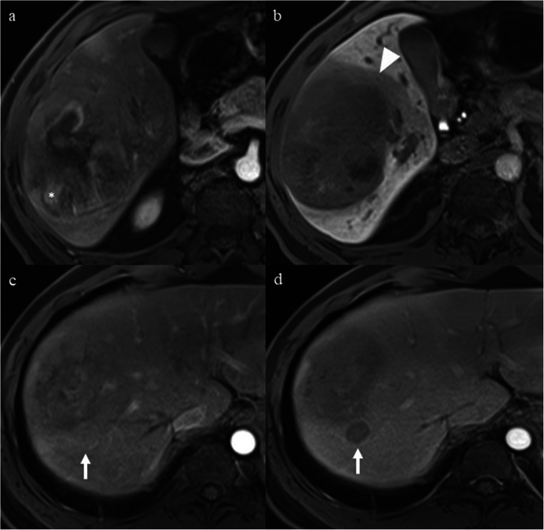
Fig. 4.
A 38-year-old man with a 10.0-cm poorly differentiated LR-5 HCC in hepatic segment V-VIII and serum AFP level of 5.06 ng/mL. a Mass shows non-rim hyperenhancement, nodule-in-nodule (asterisk), and mosaic architecture on late arterial phase. b Mass shows hypointensity, non-smooth tumor margin, and peritumoral hypointensity (arrowhead) on hepatobiliary phase. c A 1.7-cm satellite nodule (arrow) located in the peritumoral parenchyma shows non-rim hyperenhancement on late arterial phase. d The satellite nodule (arrow) shows non-peripheral “washout” on portal venous phase. This patient had two of the predictive MR imaging findings (peritumoral hypointensity on HBP and satellite nodule) for early recurrence and was categorized as LR-5c. Early recurrence occurred in the liver during follow-up after curative resection. The disease-free survival was 3.7 months

