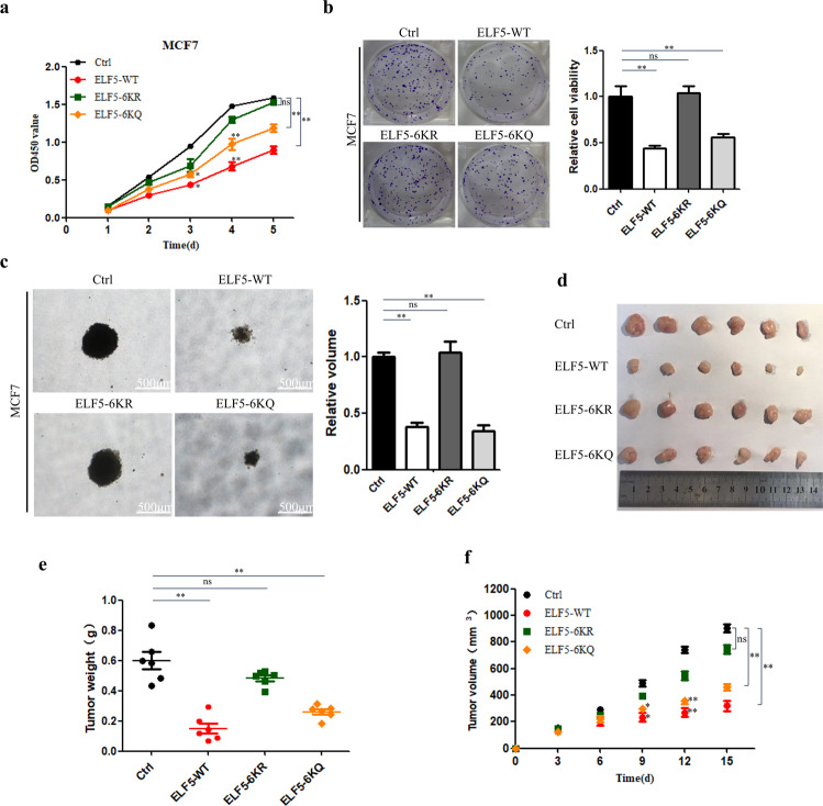
Fig. 6. Inhibition of breast cancer cell proliferation and tumorigenesis by ELF5 acetylation.
a Growth curves of MCF7 cells that stably expressed GFP-ELF5 WT, GFP-ELF5 6KR, GFP-ELF5-6KQ as measured by CCK8 assay. Control cells were transfected with just the pEGFP-C1. Data are the means ± SDs from three determinations. *p < 0.05; **p < 0.01; ns, no significance. b Colony formation of MCF7 cells that stably expressed GFP-ELF5 WT, GFP-ELF5 6KR, GFP-ELF5-6KQ as measured by crystal violet staining assay. Control MCF7 cells were transfected with just the vector pEGFP-C1. Data are the means ± SDs from three determinations. *p < 0.05; **p < 0.01; ns, no significance. c Anchorage-independent growth activity of MCF7 cells as measured by soft-agar colony culture assay. Data are the means ± SDs from three determinations. *p < 0.05; **p < 0.01; ns, no significance. d Image showing the tumor formed in xenograft mice. e, f Weight and growth of tumor induced in mice by subcutaneously implanted MCF7 cells (5 × 10⁶ cells injected per mice) that stably expressed GFP-ELF5 WT, GFP-ELF5 6KR, GFP-ELF5-6KQ. Control MCF7 cells were transfected with just the vector pEGFP-C1. Data are the means ± SDs from three determinations. *p < 0.05; **p < 0.01; ns, no significance.

