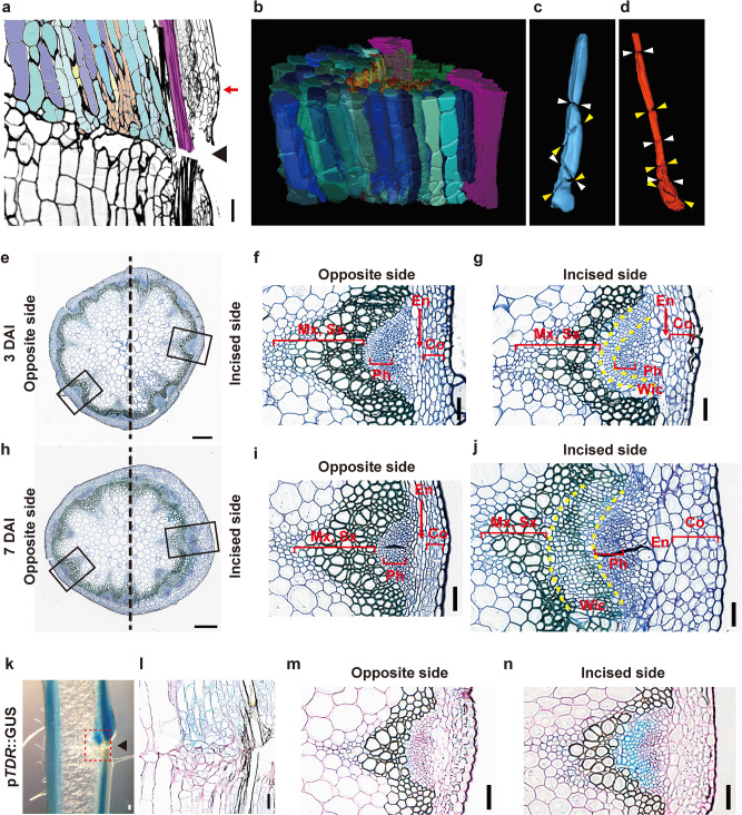
Fig. 1. Histological observation of incised flowering stem.
(a–d) Longitudinal serial sections of WT flowering stem at 4 DAI. Cells are colored by tissue as follows: pith (blue tones), parenchyma cell of protoxylem (yellowish-orange tones), secondary xylem (green), and interfascicular fiber (purple). a One of the serial sections. The arrowhead indicates the incision. b–d 3D model reconstructed from the upper region of the incision and extracted to strands of pith (c) and parenchyma in protoxylem (d) derived from one cell. The pair of arrowheads indicate the position of cell division. Thin cross-sections were located in the upper region of the incision (position of red arrow in a) at 3 (e–g) and 7 DAI (h–j). e, h Opposite and incised sides are shown on the left and right, respectively. Scale bars = 500 μm. The magnified images of the black rectangular area are shown in f, g, i, and j. The tissue between two yellow-dashed lines indicates Wic wound-induced cambium. Mx metaxylem, Sx secondary xylem, Ph phloem, En endodermis, Co cortex. k–n Histochemical analysis of pTDR::GUS in incised flowering stem. k Freehand longisection was prepared from stem at 7 DAI. l Thin longisection corresponded to the red rectangular area shown in k. m, n Thin cross-sections were prepared from the upper region of the incision at 3 DAI. Cell walls with red color were counterstained by periodic acid-Schiff. Scale bars = 100 μm.

