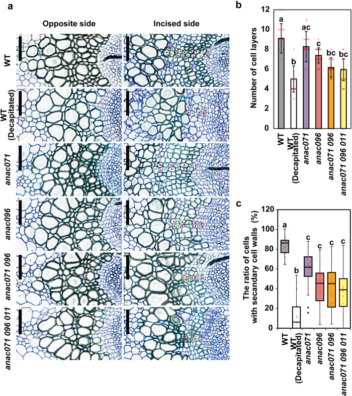
Fig. 4. Histological observation of the secondary xylem in incised stems of anac mutants.
a Thin cross-sections of vascular tissue at the upper end of the incision at 7 DAI. Left, opposite side; right, incised side. Layers of periclinally divided cells were counted as red and green dots on the incised side (bottom). Scale bars = 100 μm. b Bars are the mean number of cell layers at 7 DAI (n = 18 from 9 plants). Different letters indicate statistically significant differences (P < 0.05; Tukey’s test). c Boxplots showing the ratio of cells with secondary cell walls in the cell layer. Statistical analysis was performed using the Steel–Dwass test (P < 0.05).

