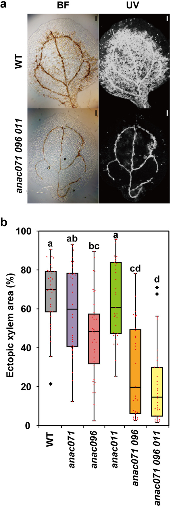
Fig. 5. Morphology of cotyledons under VISUAL assay.

a Observation of ectopic xylem vessel elements in WT and anac071 096 011 triple mutant at 4 DOC. Left, bright-field images; right, UV autofluorescence images. Scale bars = 200 μm. b Boxplots showing the percentage of ectopic xylem area in WT and anac mutants (n = 36 for WT, 34 for anac071, 35 for anac096, 33 for anac011, 31 for anac071 096, and 33 for anac071 096 011, respectively). Diamond marks indicate outliers. Statistical analysis was performed using the Steel–Dwass test (P < 0.05).
