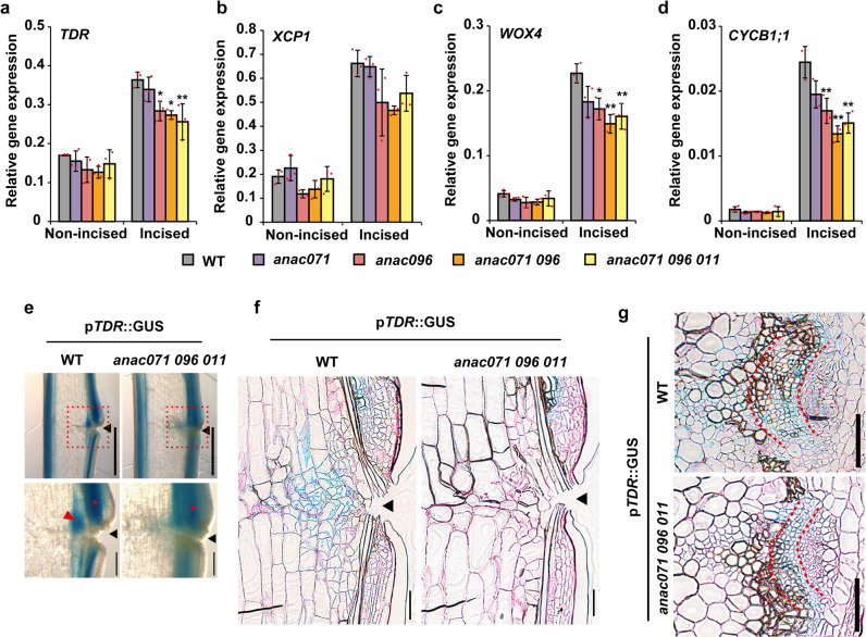
Fig. 7. Comparison of wound-induced cambium between WT and anac mutants.
a–d The relative expression levels of TDR/PXY, XCP1, WOX4, and CYCB1;1 in flowering stems were analyzed at 3 DAI. The means ± SD of triplicate batch experiments are shown. Statistical analysis was performed using Dunnett’s test, in comparison with WT (*p < 0.05). e–g Histochemical pTDR/PXY::GUS staining in the incised flowering stems of the WT and the anac071 096 011 triple mutant background at 7 DAI. e Freehand longisections. Scale bars = 1 mm. The regions in red dotted-line boxes are shown at higher magnification (bottom, Scale bars = 200 μm). Red arrowhead, wound-induced cambium of extravascular region; red asterisks, wound-induced cambium of intravascular region; black arrowheads, incised site. f Thin longisections located between vascular bundles. g Vascular bundles in the upper region of the incision. The area between two red dashed lines indicates wound-induced cambium of intravascular region. Cell walls with red color were counterstained by periodic acid-Schiff. Scale bars = 100 μm.

