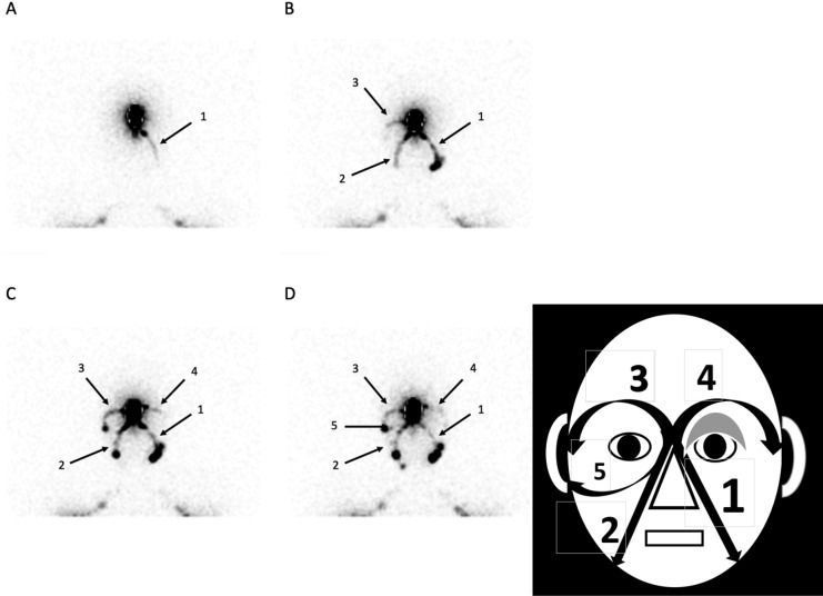
Figure 2.
This patient came with an edema at the level of the left eyelid reported as present since birth. His medical history revealed familial genetic disease (mucoviscidosis) and a malformation of the ductus thoracicus on his bipedal lymphoscintigraphic exam (no lower limb edema). His exam shows (see picture on the right and the scintigraphic images on the left) the orderly appearance of the left paranasal drainage (picture A, arrows 1), then of the right one (picture B, arrows 2) with the right supra-ocular one (picture B, arrows 3), of the left supra-ocular one (picture C, arrows 4). At the end of the exam (picture D), the right infra-ocular lymphatic drainage may be seen internal to the right preauricular lymph node (arrow 5).

