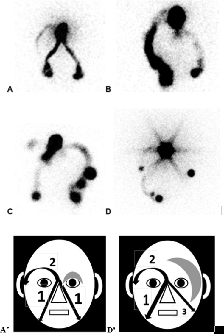
Figure 4.
Picture (A) (scintigraphic view) and (D’) (schematic presentation of the result): in this case referred for isolated edema at the level of the left eyelid (gray zone), the paranasal lymphatic drainages appear normally (arrows 1) while the appearance of the supra-ocular LV is delayed (arrow 2) and while the left supra-ocular lymphatic drainage could not be demonstrated. The 1ary origin of this abnormality was suggested by the demonstration of one associated malformation of the ductus thoracicus by a bipedal lymphoscintigraphy. Picture (B) In this case, the patient had injections of Botulinum Toxine at the level of her face. She complained of edema at the level of the eyelids but more pronounced on the left in the upper part and on the right in the external part. In her case, only the right supra ocular and left para nasal drainages could be demonstrated. Picture (C) In this patient who was referred with edema at the level of the two eyelids but more pronounced at the inferior part of the right one and who had presented right otitis, the left paranasal lymphatic drainage is not seen, the right supra ocular is not complete and the right para nasal drainage is unusually hyperactive in its upper and apical part. Picture (D) (scintigraphic view) and (D’) (schematic presentation of the result): this patient had congenital micro-cystic lymphangiomatous lesions at the level of her left edematous hemiface (gray zone). The exam showed normal right para nasal lymphatic drainage (arrow 1) and delayed supra-ocular lymphatic drainage (arrow 2) but no supra ocular on the left side and one abnormal left infra ocular drainage reaching intra-parotid LN (arrow 3).

