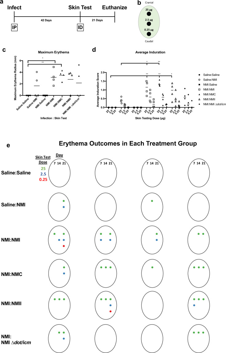
Fig. 7. Skin testing reveals the contribution of LPS and the T4BSS of C. burnetii in post-vaccination hypersensitivity.
a Animals were infected intraperitoneally (IP) with 106 NMI, or mock infected with saline, allowed to rest for 42 days, then intradermally (ID) injected (skin tested) with 25, 2.5, and 0.25 μg fixed C. burnetii or saline b Animals were then monitored for 21 days and euthanized. Erythema was measured at each skin testing site and is represented by maximum erythema radius (mm) at any day or skin testing dose c Average induration over the entire monitoring period is presented in (d). Overall erythema outcomes for each animal in each treatment group is presented in (e); both day of measurement (columns) and skin testing site dose (row) are represented. A colored dot represents measurable erythema at the corresponding day post-skin test and skin test lesion site. Maximum erythema and average induration were evaluated based on differences in group means (horizontal bar) with associated confidence intervals. For each reported comparison, p-values were computed at the α = 0.05 level for two-sided two-sample t tests, allowing for unequal variances between groups. No adjustment was made for multiple comparisons due to the sample sizes (n = 4) and the descriptive nature of the study. *p ≤ 0.05 and **p ≤ 0.01.

