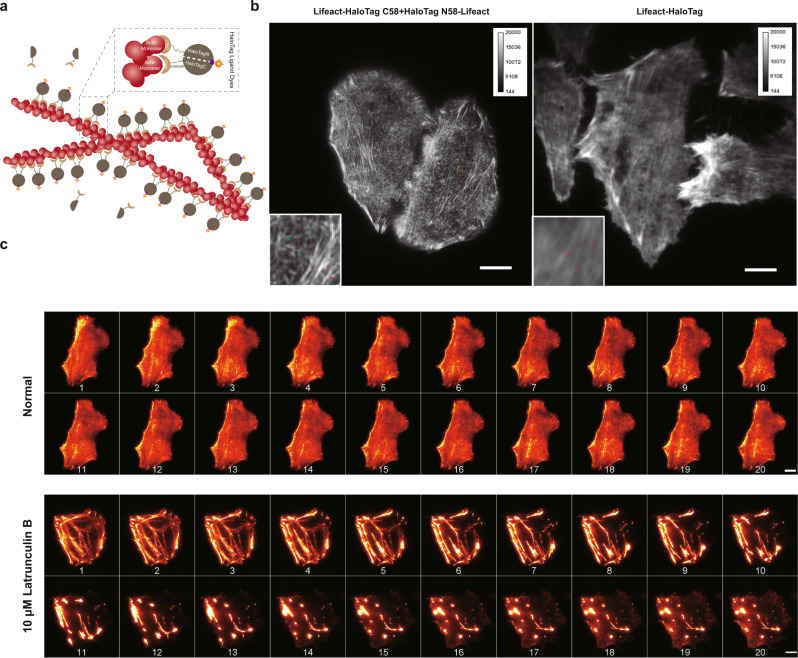
Fig. 6. Background-free imaging of actin filaments in live cells using TagBiFC.
a Schematic of actin filament imaging using BiFC-Tag. The two split halves of HaloTag were both fused with F-actin-binding peptide lifeact (Lifeact-HaloTag C58+HaloTag N58-Lifeact). Only two halves that bind to two adjacent actin units of the filaments and reconstitute an intact HaloTag can be imaged under microscopy. b Comparation of F-actin labeling results by BiFC-Tag (left) and HaloTag (right). Insets show the box regions in the images. Red arrow heads indicate the thick stress fibers that can be labeled using both methods while the green arrow heads indicate the thin filaments that can only be labeled using TagBiFC. Scale bars: 5 μm. c BiFC-Tag allows long-term imaging of F-actin dynamics under normal or Latrunculin treatment conditions. Representative snapshots were displayed, and time post treatment is shown under each snapshot. Scale bars: 5 μm.

