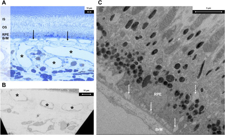
Figure 3.
Histologic findings following systemic adrenaline (group 2). (A) Light microscopy section with toluidine blue stain at magnification × 40 demonstrating dilated choroidal venules (indicated with black asterisks) and choriocapillaris (indicated with solid black arrows). (B) Electron microscopy section at magnification × 440 demonstrating dilated choroidal venules (indicated with black asterisks). (C) Electron microscopy section at magnification × 1900 demonstrating an intracytoplasmic vacuole at the basal aspect of RPE cells (indicated with interrupted white arrow) and enlarged basal infoldings (indicated with solid white arrows). IS, inner segment; OS, outer segment; BrM, Bruch's membrane.

