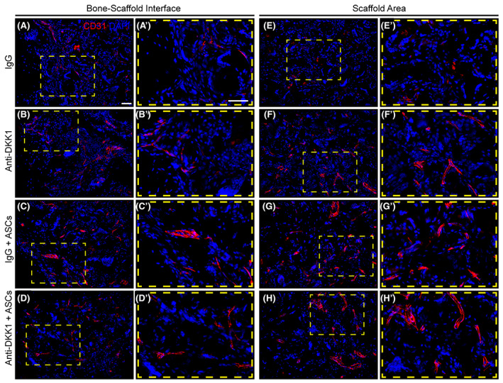
FIGURE 7.

Vascular ingrowth is promoted by both ASC therapy and DKK1 neutralization. Defects were treated with ASC seeded scaffolds or acellular control scaffolds. Animals were treated with anti‐DKK1 or IgG control (15 mg/kg, SC, twice weekly). CD31 immunohistochemical staining of bone‐scaffold interface (A‐E), and within the implant site (E‐H). High magnification insets show in detail of vascular distribution (A′‐H′). CD31+ endothelial cells appear red, while DAPI nuclear counterstain appears blue. All analyses performed at 8 weeks postimplantation. White scale bars = 50 μm. ASCs, adipose‐derived stem cells. DAPI, 4′,6‐diamidino‐2‐phenylindole
