Figure 5.
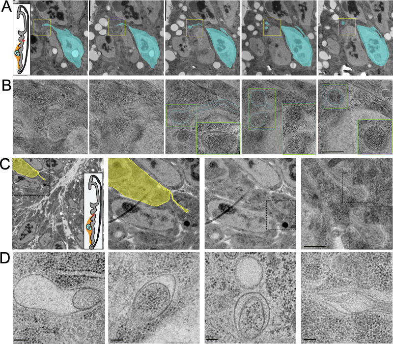
Disc cytonemes extend into invaginations near the apical and basal surfaces. (A) A series of five TEM image montages showing a disc epithelial cell (cyan) projecting a cytoneme that ends in a membrane invagination of a neighboring disc cell. The region imaged is shown as an asterisk on the disc diagram. Part of the target cell’s apical surface and microvilli are seen in the upper left corner. (B) The yellow box represents the area montaged at higher magnification where the path of the cytoneme in the invagination is seen. Inside the invagination, the cytoneme branches off into two terminal varicosities. (C) A TEM image shows a disc epithelial cell (yellow) projecting a cytoneme from its lateral surface, which ends in an invagination of the membrane of a neighboring disc cell near its basal surface. The area imaged is in the fold indicated on the disc diagram. Progressive enlargements of the boxed regions are shown from left to right. The middle two images are the same with and without segmentation of the cytoneme-projecting cell. (D) TEM images of four separate endings of cytonemes within invaginations of target cell membranes, showing the variability in the size of the space between the end of the cytoneme and target cell membranes. Scale bars: 500 nm (A and B), 2 µm (C panel 1), 500 nm (C panel 4), and 100 nm (D).

