Figure 6.
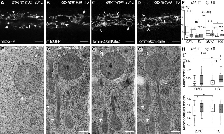
HS-induced mitochondrial fragmentation is dependent on DRP-1. (A–D) Confocal images of the mitochondria in the epidermis of drp-1 animals visualized with the matrix-located mitoGFP (A and B) or outer membrane TOMM-20::mKate2 (C and D) at 20°C (A and C) or after HS (B and D). Abnormally shaped mitochondria are classified as enlarged (large arrow), clustered (arrowhead), and filament-like (small arrow). For comparison with wild type, see Fig. 2, D, E, I, and J. (E) Boxplots of the mitochondrial shape descriptors form factor (FF) and aspect ratio (AR) using mitoGFP. Seven or eight animals were analyzed, and >700 mitochondria were measured per animal. ***, P < 0.001, Wilcoxon test. (F–G″) EM pictures of mitochondria in the epidermis of drp-1 animals at 20°C (F) or after HS (G). G, G′, and G″ are serial sections of stress mitochondria containing dark occlusions and displaying enlarged (large arrows), clustered (arrowhead), and filament-like (small arrow) shapes. For comparison with wild type, see Fig. 1, A and B. (H) Measurements of the area and the roundness of mitochondria in the epidermis were performed on EM pictures. Boxplots; n = 40, 65, 37, 51; *, P < 0.05; ***, P < 0.0005, Kruskal-Wallis test. The scale bars are 10 µm (A–D) or 0.5 µm (F and G).

