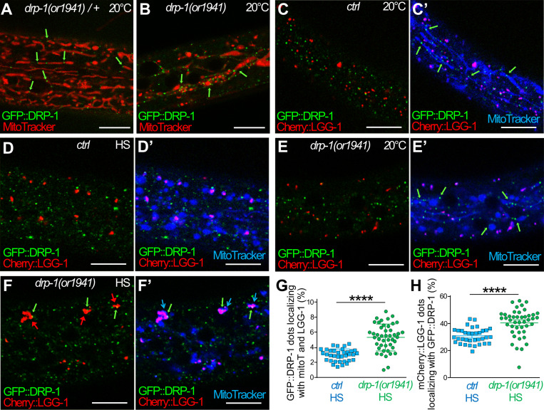
Figure 8.
The dysfunction of DRP-1 during aHS induces the accumulation of autophagosomes at mitochondrial fission sites. (A and B) Confocal images of GFP::DRP-1 (green) and MitoTracker (red) in the epidermis of heterozygous control (A) and homozygous drp-1(or1941) mutants (B). drp-1(or1941) is an in-frame insertion of GFP at the drp-1 locus that results in an abnormal mitochondrial network (blebs in B), indicating that GFP::DRP-1 is correctly localized (green arrows) but is not functional. This particular GFP allows visualizing the presumptive mitochondrial fission points in a context-phenocopying drp-1 mutant (B). (C) Another transgenic GFP::DRP-1 strain where the drp-1 locus wild type is used as a positive control. (C–F) Confocal live imaging of the epidermis of GFP::DRP-1 in control (C and D) and drp-1(or1941) mutant animals (E and F) at 20°C (C and E) and after HS (D and F). The mitochondria are stained with MitoTracker DeepRed (blue), and autophagosomes are visualized with mCherry::LGG-1 (red). C′, D′, E′, and F′ show three colors merging. (C) In control animals at 20°C, the GFP::DRP-1 puncta are associated to tubular mitochondria but not to mCherry::LGG-1 dots (C′). (D) Upon aHS, most of the GFP::DRP-1 puncta are not localizing with mitochondria (B) or with mCherry::LGG-1 dots. (E) In drp-1(or1941) mutant at 20°C, the presumptive mitochondrial fission points (GFP puncta) are associated with blebs or filamentous mitochondria but not with mCherry::LGG-1 dots (E′). (F) After aHS of drp-1(or1941) mutants, the clustering of autophagosomes (red arrows pointing to mCherry::LGG-1) is associated with presumptive mitochondrial fission points (green and blue arrows in F′). (G) Quantification of the colocalization of GFP::DRP-1 puncta with MitoTracker (mitoT) and mCherry::LGG-1 in the epidermis of control and drp-1(or1941) animals after aHS (mean and SEM, n = 35, 44; ****, P < 0.0001, t test with Welch correction). (H) Quantification of the colocalization of mCherry::LGG-1 puncta with GFP::DRP-1 in the epidermis of control and drp-1(or1941) animals after aHS (mean and SEM, n = 35, 44; ****, P < 0.0001, t test with Welch correction). The scale bars are 10 µm.

