Figure S5.
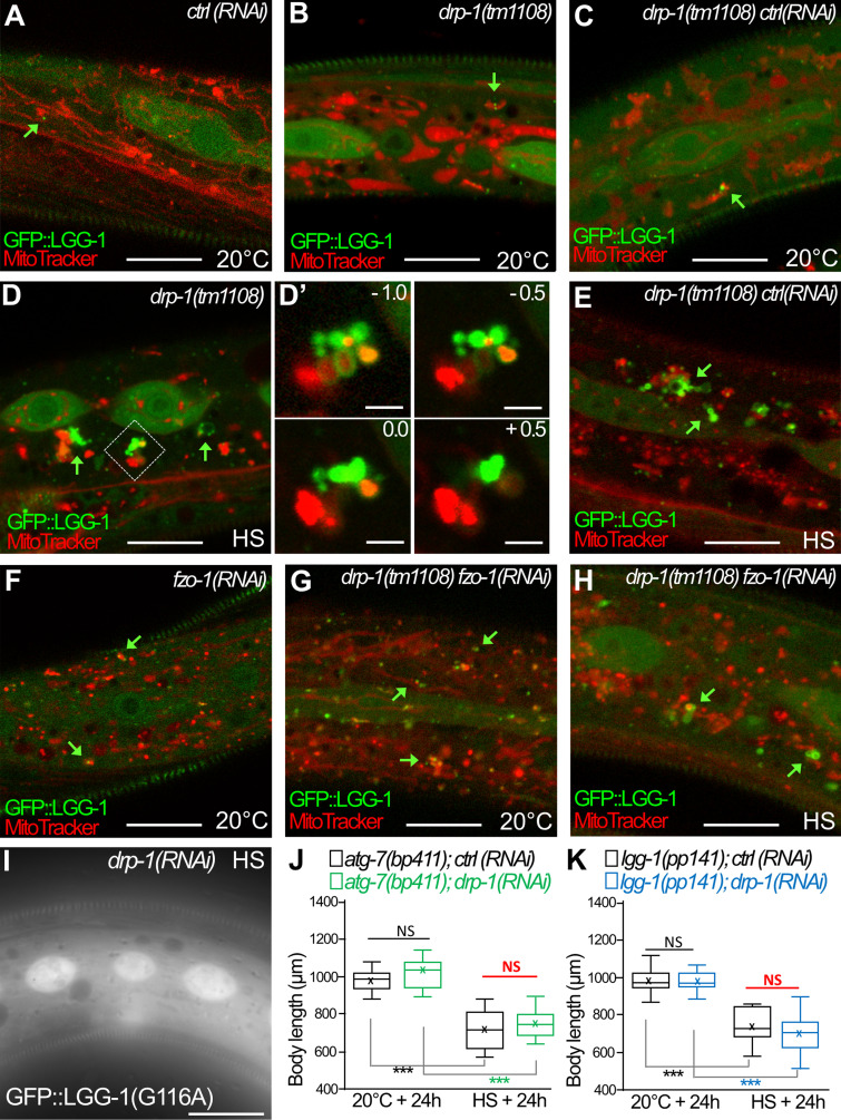
The clustering of autophagosomes at mitochondria is maintained in drp-1;fzo-1 animals after aHS (complementary to Fig. 7). (A–H) Confocal images of autophagosomes (GFP::LGG-1; green) and mitochondria (MitoTracker; red) in the epidermis of animals at 20°C (A–C, F, and G) and after aHS (D, E, and H). Green arrows point to autophagosomes in contact with mitochondria. In drp-1 animals (A–D), the aHS induces an accumulation of autophagic structures intermingled with mitochondria. Insets in D′ are 0.5-µm Z-series corresponding to the dotted square in D. The depletion of FZO-1 induces a fragmentation of the mitochondrial network in control animals (F) but partially restores a tubular mitochondrial network in drp-1(tm1108) animals (G). In drp-1(tm1108);fzo-1(RNAi) animals, upon aHS, the GFP::LGG-1 forms large clusters in contact with mitochondria. (I) The nonlipidated GFP::LGG-1(G116A) does not form puncta or clusters after HS in drp-1 animals. (J and K) The depletion of DRP-1 does not further increase the developmental delay of autophagy mutants (atg-7, lgg-1) induced by aHS. Single mutants with ctrl(RNAi) or dpr-1(RNAi) were maintained at 20°C or submitted to aHS and measured after 24-h recovery (boxplots, n > 30; ***, P < 0.0001, Kruskal-Wallis test). The scale bars represent 10 µm or 2 µm (D′).

