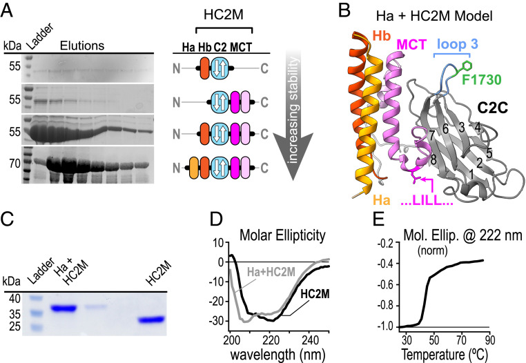
Fig. 3.
C2C together with N-terminal helices and the MCT form a membrane-binding protein domain. (A) Protein gels of C2C domain elutions (Left) and domain cartoons (Right) for several constructs differing in the amount of neighboring protein included in the expression construct assuming a type-I topology for the C2 domain. (B) A proposed model for HC2M structure (reference SI Appendix for details). Protein gel (C) and CD spectra (D) of recombinant worm Hb-C2C-MCT (black) and Ha-Hb-C2C-MCT (gray) following cleavage from maltose-binding protein. The minimal stable domain (Hb-C2C-MCT) is referred to as HC2M. (E) Thermal melting plot of WT HC2M (black).

