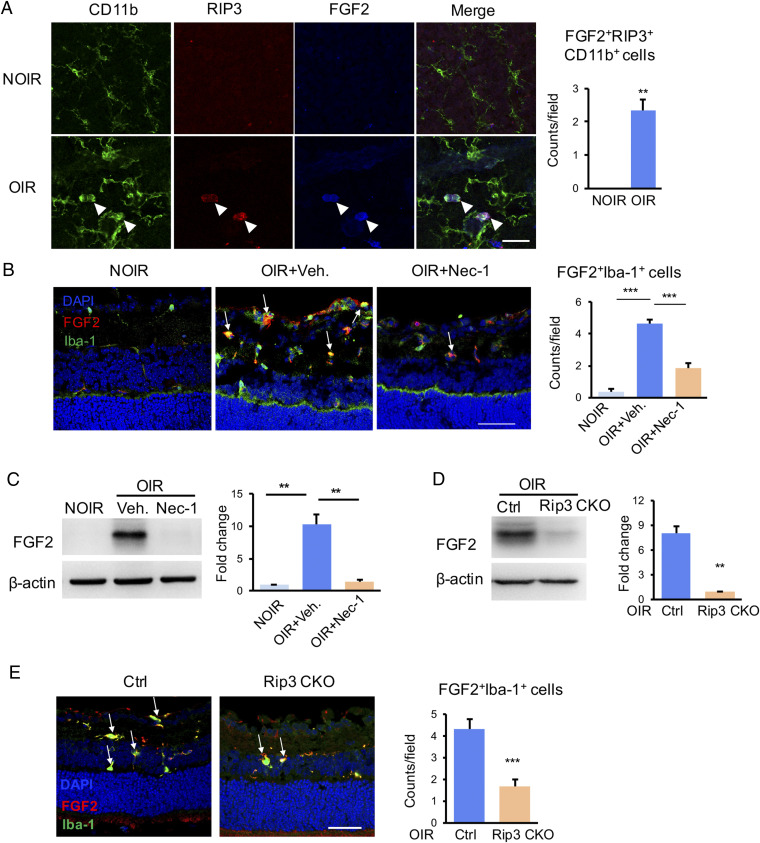
Fig. 4.
Improved FGF2 production in necroptotic microglia in vivo. (A) Immunofluorescence staining in retinal whole mounts revealed strong staining of FGF2 in RIP3+CD11b+ necroptotic microglia from OIR retina (white arrowheads), while lack of FGF2 and RIP3 expression in the NOIR control. n = 3 retinae, 6 images/retina. (B) The FGF2+Iba-1+ microglia were increased in OIR, which was abrogated by Nec-1. n = 3 eyes, 6 sections/eye. (C) The Western blot result showed that the elevated FGF2 in OIR were inhibited significantly in the Nec-1–treated ones. n = 6 retinae. (D) The FGF2 expression was markedly suppressed in the OIR retina from Rip3 CKO mice in comparison to controls. n = 6 retinae. (E) The FGF2+Iba-1+ microglia were also highly reduced in Rip3 CKO-OIR retina. n = 3 eyes, 6 sections/eye. (Scale bars: 50 µm in A, B, and E.) **P < 0.01; ***P < 0.001.

