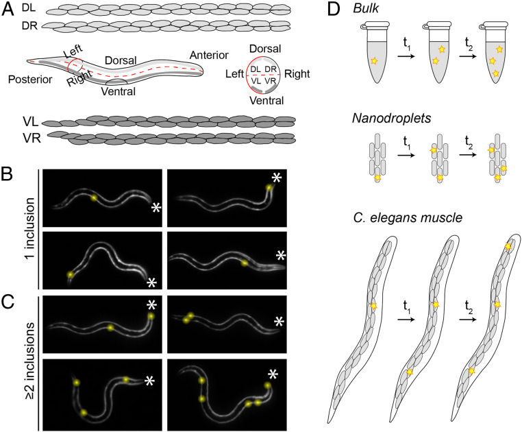
Fig. 2.
Q40-YFP aggregation occurs stochastically in individual muscle cells. (A) Illustration showing the distribution of the 95 body wall muscle cells in adult C. elegans. Four bundles of muscle cells run along the posterior–anterior axis. The animals typically crawl on their left or right side on solid media, resulting in a superposition of the two dorsal and the two ventral bundles (Middle Left). A schematic view from the anterior side shows the localization of the four bundles in the different quadrants (Middle Right). Cell shapes were drawn based on http://wormatlas.org. (B) The 32-h-old animals display their first inclusion at any position along the posterior–anterior axis. The asterisks mark the anterior of the animals; inclusions are highlighted by yellow spheres for better visualization. (C) The 32-h-old animals with two or more inclusions support the notion that nucleation is initiated stochastically in individual cells, in the absence of a pattern of spatial propagation from the first to subsequent inclusions. (D) Cartoon showing the occurrence of aggregation events (yellow stars) in bulk, nanodroplets, and in C. elegans muscle cells. In a typical test tube reaction (Top), all aggregation events take place within the same continuous volume. In nanodroplets (Middle) and C. elegans muscle tissue (Bottom, only one bundle of muscle cells is shown for clarity), the total volume is divided over multiple small volumes, in which the probability of nucleation is low. As a consequence, aggregation will be initiated in different droplets or cells at different points in time. Three aggregation events are shown in each system for clarity, but in reality the number of nucleation events in a given period of time is proportional to the size of the reaction vessel.

