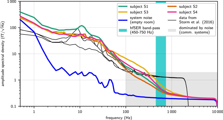
Fig. 1.
Amplitude spectral density of resting-state recordings compared with system noise without subject. The system white noise level (blue line) in the empty magnetically shielded recording room was detected at 0.18 , approximately one order of magnitude below the noise level of standard MEG systems. The gray shaded area indicates the range of frequencies and signal amplitudes dominated by system noise in case of a standard MEG system. Above 200 Hz (and extending through and even beyond the subsequently analyzed hfSER frequency band [cyan shading]), the ultralow-noise MEG still continued to show the physiological 1/f-spectral decay in resting-state recordings for all subjects (color-coded lines; cf. Inset) that in standard recordings would be hidden in system noise. The minute offset between system noise level and resting-state recordings persisting in the hf range (especially approximately >5,000 Hz) is mainly due to the natural thermal body noise (16, 21, 22). For a direct comparison, reanalyzed data from the immediate technological predecessor system (23) are shown featuring a high-frequency white magnetic field noise lower than commercial systems, but still obscuring biological activity at frequencies above 300 Hz (black lines; three subjects). The signal drop at 2,500 Hz is caused by low-pass filtering due to a lower sampling rate of the recordings (5,000 samples/s).

