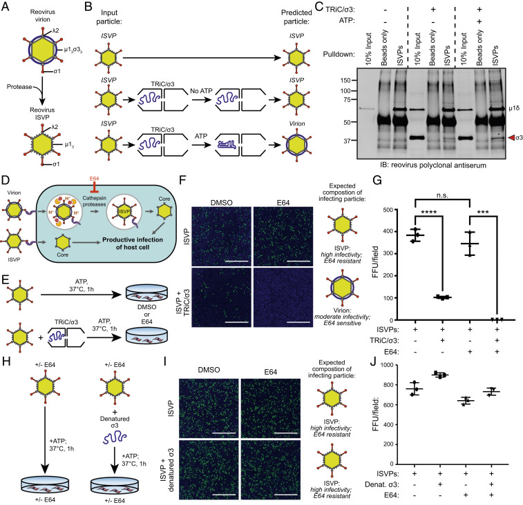
Fig. 7.
TRiC/σ3 recoats ISVPs producing biologically active mature reovirus particles. (A) Schematic of reovirus virion and ISVP resulting from proteolytic disassembly of virions. (B) Workflow of ISVP recoating experiment. (C) Immunoblot of immunoprecipitated ISVPs incubated in the presence or absence of TRiC/σ3. (D) Schematic illustrating the reovirus virion and ISVP entry pathways and inhibition of virion disassembly by E64. (E) Workflow to assess ISVP infectivity when incubated with TRiC/σ3. (F) Immunofluorescence images of HeLa cells cultured in DMSO or E64 (200 μM) and inoculated with ISVPs (Top) or ISVPs incubated with TRiC/σ3 (Bottom) and stained with DAPI (blue) and a reovirus-specific antiserum (green). (Scale bar: 1 mm.) (G) Quantification of fluorescent focus units (FFUs) per imaging field from the experiment in F. (H) Workflow to assess the infectivity of ISVPs incubated with denatured σ3. (I) Immunofluorescence images of HeLa cells incubated with DMSO or E64 and inoculated with ISVPs (Top) or ISVPs incubated with denatured σ3 (Bottom) and stained with DAPI (blue) and a reovirus-specific antiserum (green). (Scale bar: 1 mm.) (J) Quantification of FFUs per imaging field from the experiment in I. For quantifications, results shown are the mean ± SD of a single experiment conducted in triplicate and are representative of three independent experiments (***P < 0.001; ****P < 0.0001; unpaired two-tailed t test; n.s., not significant).

