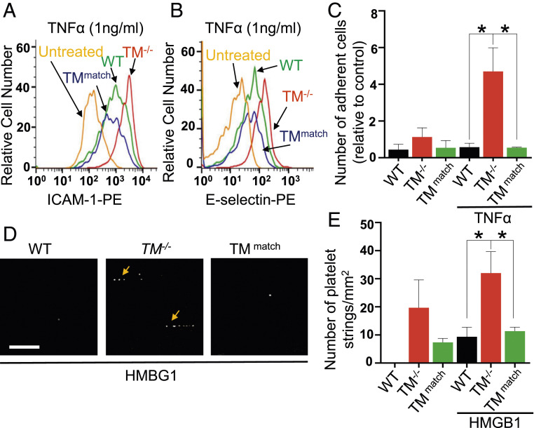
Fig. 3.
Lentivirus-mediated reexpression of TM in TM−/− cells inhibits TNF-α-induced adhesion of HL-60 cells and HMBG1-induced platelet string formation on TM−/− cells. (A) Cell surface levels of ICAM-1 and (B) E-selectin in WT, TM−/−, and TMmatch cells, treated with TNF-α (1 ng/mL) for 4 h, were measured by flow cytometry. TM−/− cells were used as untreated controls. (C) Fold change in the adhesion of HL-60 cells on WT, TM−/−, and TMmatch cells, stimulated with TNF-α (1 ng/mL) for 4 h under flow conditions per field of the view (firmly adherent cells were quantified). (D) WT, TM−/−, and TMmatch cells were incubated with HMGB1 (20 nM) for 18 h and then 5-chloromethylfluorescein diacetate–labeled platelets were perfused in a flow chamber. Representative images showing platelet string formation captured on WT, TM−/−, and TMmatch cells. (Scale bar, 25μm.) (E) Quantitation was measured by counting the average number and length of strings per field in different groups. The average number of platelet strings per field was determined as ≥3 platelets aligned in the direction of the flow. Data are mean ± SEM (n = 3). One-way ANOVA: *P < 0.05.

