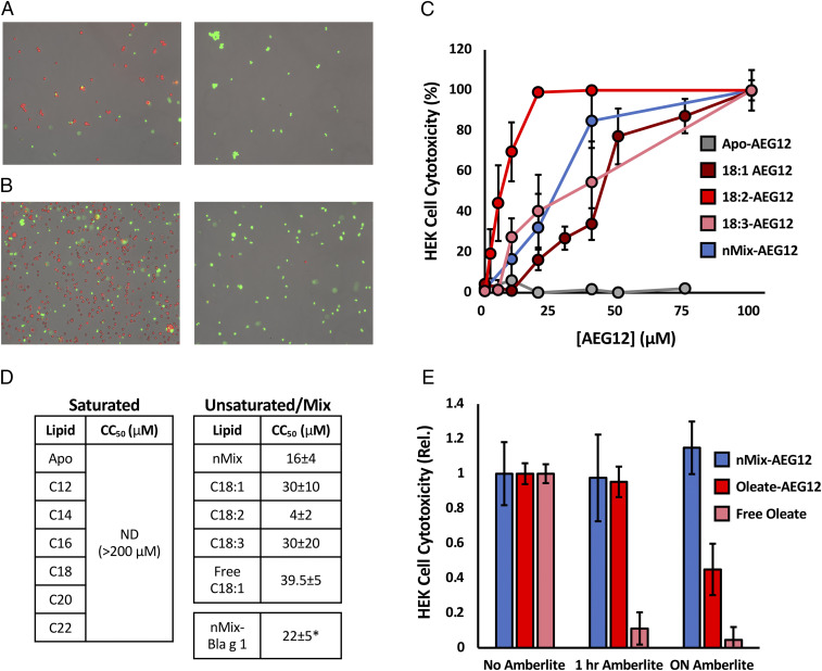
Fig. 3.
AEG12 cytotoxic activity in mammalian cells is restricted to unsaturated fatty acid ligands. (A) Microscopy images obtained at 10× for HEK cells treated with PBS (Left) or 2% Triton 100 (Right) representing negative and positive controls, respectively. Viable (live) and dead cells are identified using the AO/PI stain system and are colored red and green, respectively. (B) Cells treated with 40 μM Apo-AEG12 (Left) or nMix-AEG12 (Right) and imaged as in A. HEK cell cytotoxicity, defined as the proportion of dead cells to total cells, following treatment with varying concentrations of AEG12 shown in C. 50% cytotoxicity (CC50) values for AEG12 loaded with various saturated and unsaturated cargoes are shown in D. ND denotes fatty acid cargoes for which no detectable lysis was observed at any of the concentrations tested. (E) Decrease in cytotoxicity of nMix-AEG12 (blue), Oleate-AEG12 (red), or free oleate alone (pink) following 1 h and overnight incubation with Amberlite resin relative to an untreated (no Amberlite) control. * denotes values obtained from two independent trials.

