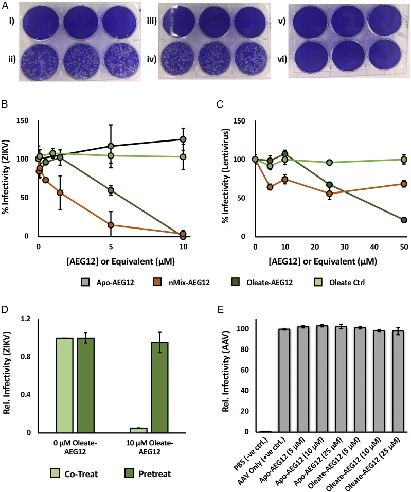
Fig. 4.
AEG12 inhibits Zika virus, lentivirus infection. (A) Representative plaque assay plate showing the formation of Zika virus (ZIKV Paraiba 2015)-induced plaques on confluent Vero cells (stained blue). Rows i) and ii) depict plates treated with no virus, or untreated virus, representing the negative and positive controls, respectively. Treating the virus with 10 μM Apo-AEG12 for 30 min did not significantly reduce infectivity (iv), while treatment with 10 μM nMix-AEG12 completely inactivated the virus (vi). Treatment of the cells with identical concentrations of Apo-AEG12 or nMix-AEG12 alone did not significantly hinder proliferation in the absence of virus, as shown in iii) and v), respectively. A plot of the relative number of viral plaques relative to AEG12 concentration yields an inhibition curve (B) from which approximate IC50 values can be obtained. Note that the concentration of free oleate employed in the oleate control is eight times that of AEG12 in order to account for the observed 8:1 binding stoichiometry. The inhibition curve of AEG12 against the enveloped lentivirus is shown in C. Preincubating target cells with Oleate-AEG12 prior to infection by ZIKV (pretreat) yields no protective effect (D). Infectivity of ZIKV incubated with the equivalent concentration of Oleate-AEG12 as described in B (cotreat) shown for comparison. (E) Infectivity of AAV is undiminished after treatment with Apo- and Oleate-AEG12.

