Fig. 5.
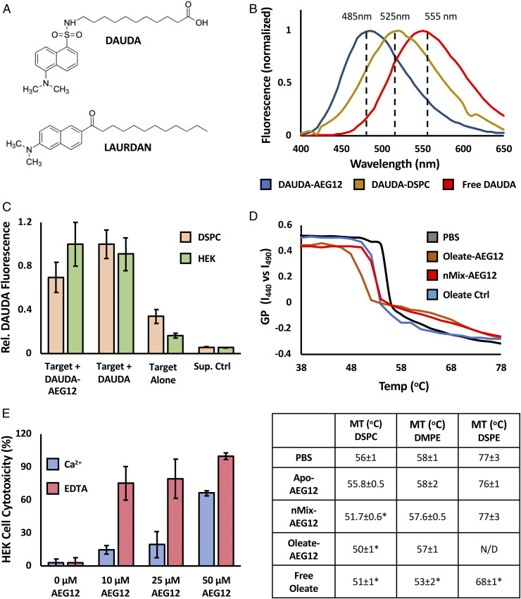
Cargo delivery by AEG12 selectively disrupts PC bilayers. (A) Chemical structure of DAUDA and LAURDAN fluorophores. (B) Normalized fluorescence intensity of DAUDA complexed with AEG12 (blue), DSPC (yellow), or in aqueous solution (Red) showing the effect of local environment on fluorescence maximum. (C) Transfer of DAUDA into DSPC vesicles (orange) or HEK Cells (green) bilayer targets following incubation with DAUDA-AEG12 or DAUDA alone followed by a three washes with PBS. Equivalent values obtained for a PBS-treated negative control (target alone) and supernatant (Sup. Ctrl) from the DAUDA-AEG12 sample shown for reference. (D) Representative melting curves showing the temperature dependence of LAURDAN GP incorporated into DSPC bilayer vesicles following incubation with 28 μM Apo-nMix, or Oleate-AEG12, or the equivalent concentration (224 μM) of free oleate. GP values were calculated from the LAURDAN emission intensity at 440 and 490 nm as discussed in the methods section. The temperature at the inflection point represents the phase transition temperature, values of which are summarized below for DSPC and DSPE vesicles treated with 28 μM or 40 μM AEG12, respectively. Conditions which differ significantly from the PBS control as determined via Student’s t test assuming equal variance are denoted with *. (E) Cytotoxicity of Oleate-AEG12 against HEK Cells in the presence of 1.3 mM Ca2+ (blue) or 2.5 mM EDTA (red) showing the inverse dependence of cell lysis on extracellular calcium and by extension the cell membrane repair pathway.

