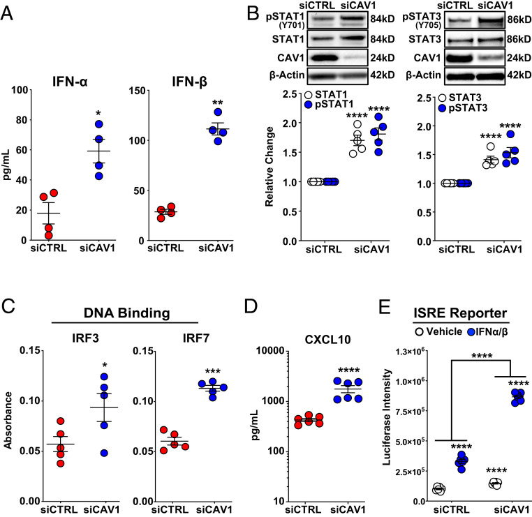
Fig. 3.
Constitutive activation of IFN signaling due to CAV1 loss-of-function in primary human PAECs. (A) IFN-α and IFN-β, measured by ELISA, were significantly higher in conditioned media 48 h after CAV1-silencing compared to siCTRL-transfected PAECs. Data presented as mean ± SE. (B) Whole-cell lysate immunoblotting 48 h after CAV1 knockdown demonstrated a significant increase in both total and phosphorylated (activated) STAT1 and STAT3. Densitometric quantification of protein normalized to β-actin and relative to siCTRL presented as mean ± SE with representative Western blots. (C) Nuclear protein isolated from CAV1-silenced PAECs 48 h after knockdown demonstrated significantly increased IRF3 and IRF7 DNA binding compared to siCTRL-transfected cells (mean absorbance ± SE). (D) CAV1 loss in PAECs led to significant increase in CXCL10 protein. Cell supernatants collected 48 h after CAV1 knockdown. CXCL10 measured by ELISA presented as mean ± SE. (E) ISRE activity was significantly higher in CAV1-silenced human EAhy926 endothelial cells in the absence and presence of IFN-α/β stimulation. Additionally, CAV1-silenced EAhy926 cells displayed significantly greater response to IFN-α/β stimulation compared to siCTRL-transfected cells. Human EAhy926 endothelial cells were stably transfected with ISRE reporter constructs and then transiently transfected with CAV1-specific or nontargeting control siRNA. After siRNA transfection (48 h), cells were treated with IFN-α/β (10 U/mL) or vehicle alone (PBS with 0.1% BSA) for 5 h. Luciferase intensity presented as the mean ± SE. Each independent experiment used different PAEC donors. *P < 0.05, **P < 0.01, ***P < 0.001, ****P < 0.0001.

