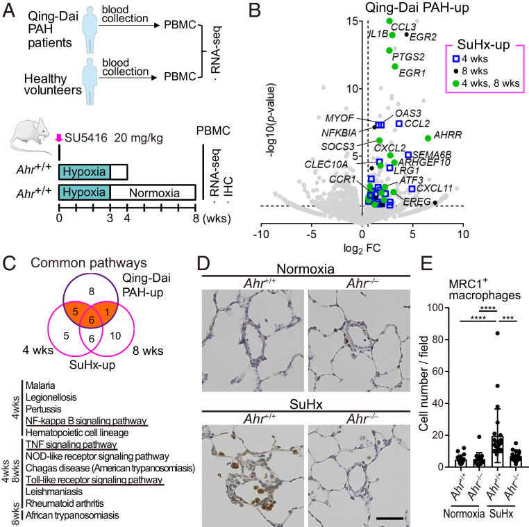
Fig. 8.
Activation of AHR induces inflammation-related genes in PBMCs of SuHx rats and Qing-Dai–induced PAH. (A) Experimental protocol for RNA-seq of PBMCs in Qing-Dai–induced PAH patients (n = 2) and PBMCs in SuHx rat lungs (n = 3 for each group). (B) Volcano plot of gene expression of PBMCs in Qing-Dai–induced PAH patients vs. HV. Genes up-regulated in PBMCs of both patients and SuHx rats at 4 and 8 wk, identified by RNA-seq experiments, are indicated by blue open squares (4 wk), black circles (8 wk), and green circles (4 and 8 wk), respectively. (C) Pathway analysis of genes up-regulated by Qing-Dai and SuHx. Orange indicates the 12 common pathways, which are shown in the lower panel. (D) Representative immunohistochemical images of pulmonary arterioles stained for MRC1 in Ahr+/+ and Ahr−/− rats. Arrows indicate MRC1+ macrophages. (Scale bar, 50 μm.) (E) Number of MRC1+ macrophages in 724 mm × 541 mm fields captured around arteries (number of tested rats: normoxia Ahr+/+: n = 3, normoxia Ahr−/−: n = 3, SuHx 8-wk Ahr+/+: n = 4, SuHx 8-wk Ahr−/−: n = 3). Values are means ± SD; ****P < 0.0001, ***P < 0.001.

