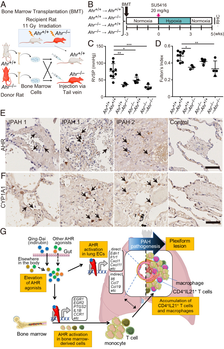
Fig. 9.
The AHR signaling pathway both in ECs and in bone marrow-derived immune cells contributes to the pathogenesis of PAH. (A) Illustrative scheme of BMT experiment using Ahr+/+ and Ahr−/− rats. Ahr+/+ or Ahr−/− BM was transferred to Ahr+/+ or Ahr−/− rats, respectively, after lethal irradiation (11 Gy). (B) Experimental protocol for RHC experiment for SuHx rats after BMT. Three weeks after BMT, SU5416 was subcutaneously administered to the rats. (C and D) Assessment of the SuHx rat model of BMT rats (Ahr+/+ BM-transplanted Ahr+/+: n = 9; Ahr+/+ BM-transplanted Ahr−/−: n = 3; Ahr−/− BM-transplanted Ahr+/+: n = 5; Ahr−/− BM-transplanted Ahr−/−; n = 4). RVSP (C), Fulton’s index (D). (E) Representative images immunostained for AHR. Arrows indicate nuclear localization of AHR in ECs (IPAH 1, Left) and in infiltrating cells in a plexiform lesion (IPAH 1, right, and IPAH 2). No nuclear localization was seen around arteries in the control (Control). (F) Representative images immunostained for CYP1A1. Arrows indicate positive signals in some ECs (IPAH 1, Left) and in infiltrating cells in plexiform lesions (IPAH 1, right, and IPAH 2). No positive staining was seen around arteries in the control (Control). (Scale bar, 50 μm.) (G) Schematic illustration of development of PAH via AHR activation. This illustration was created using Servier Medical Art (https://smart.servier.com) and BioRender (https://biorender.com). Values are means ± SD; ***P < 0.001, **P < 0.01, *P < 0.05.

