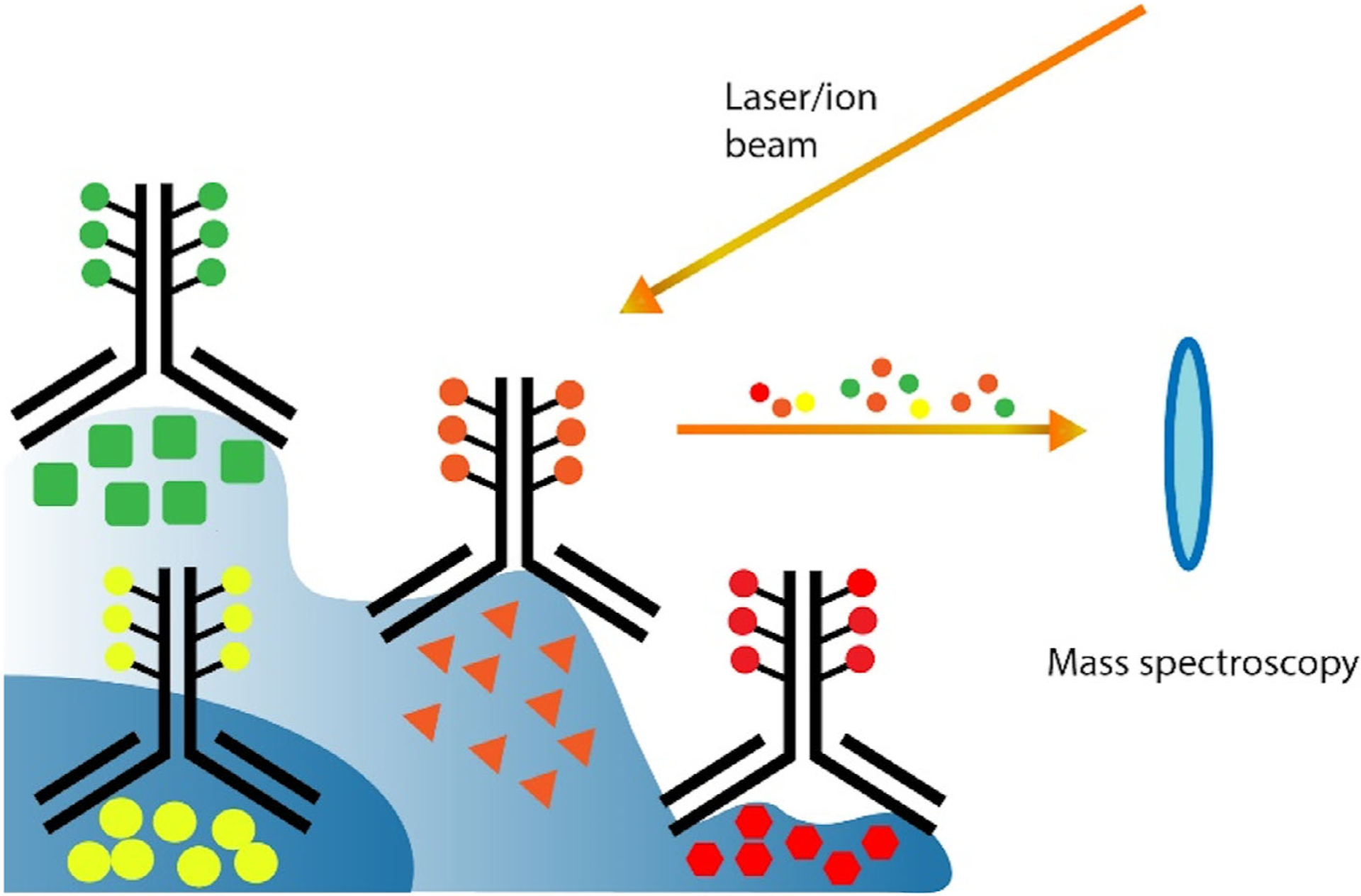
FIGURE 6.

Imaging mass cytometry and ion beam imaging. Tissues are stained with a mixture of metal isotope labeled antibodies. Then, a laser or ion beam is applied to transfer the specimen pixel-by-pixel into a mass spectrometer. The mass data of the identified metal isotopes are translated into protein abundances with computer software
