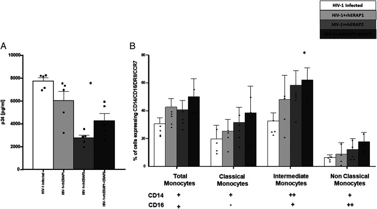
FIGURE 6.
rhERAPs stimulation favors monocyte activation and differentiation in specific subsets in an in vitro HIV-1 infection assay. (A) PBMCs from five HCs were treated with 100 ng/ml of rhERAP1, rhERAP2, or rhERAP1+rhERAP2 and in vitro HIV-infected with a R5 HIV-1Ba-L. P24 concentration was measured by ELISA in 6 d post–in vitro HIV-1–infected supernatants. Mean values and SEM are shown. *p < 0.05. (B) In the same cell cultures, 3 d post–in vitro HIV-1 infection, the percentage of total (CD14+ CD16+), classical (CD14++CD16−), intermediate (CD14++CD16+), and nonclassical (CD14+CD16++) monocytes expressing the activation markers DRII and CCR7 was assessed by flow cytometry. Mean values and SEM are shown. *p < 0.05.

