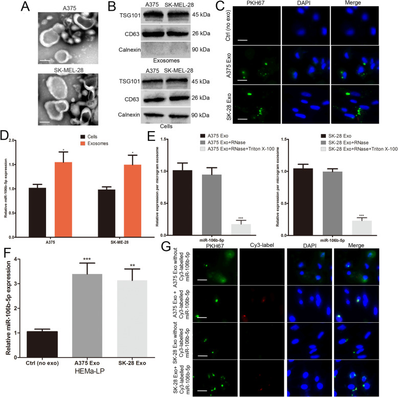
Fig. 2.
miR-106b-5p is enriched in melanoma cell-secreted exosomes and transferred to melanocytes. a Exosomes purified from culture supernatant of A375 and SK-MEL-28 cells were detected by TEM. Scale bar, 50 nm. b Western blots identified the exosomes marker proteins CD63 and TSG101, and Calnexin was used as an internal reference. c Exosomes purified from culture supernatant of A375 and SK-MEL-28 cells were labeled by PKH67. HEMa-LP cells was co-cultured with these exosomes and observed under confocal microscope, non-exosomes group was used as the negative control. Scale bar, 20 μm. d Basic miR-106b-5p levels in melanoma cells and paired exosomes were detected by qRT-PCR. e miR-106b-5p expression in exosomes, untreated or treated with RNase A and/or Triton X-100. f miR-106b-5p levels in HEMa-LP cells pre-treated with non-exosomes or indicated exosomes for 24 h were detected by qRT-PCR. g Exosomes with Cy3-labeled miR-106b-5p were added to HEMa-LP cells, fluorescence signals were detected under confocal microscope. miR-106b-5p without Cy3-label was used as the negative control. Scale bar, 20 μm. Data were expressed as the mean ± SD, *P < 0.05, **P < 0.01, ***P < 0.001

