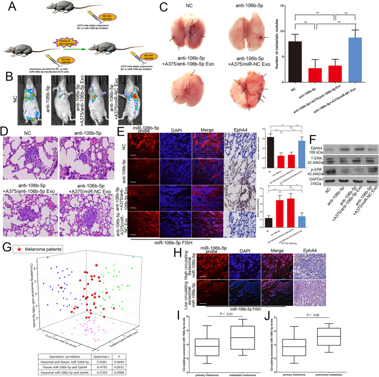
Fig. 6.
Exosomal miR-106b-5p promotes melanoma metastasis in vivo. a The schema of the animal experiment. b Representative bioluminescence images of mice after tail vein injection of stably expressing miR-106b-5p inhibitor A375 cells with or without intravenous injection of exosomes secreted by A375 cells. c The excision lung tissues in nude mice and the number of metastatic lung nodules. d Metastatic lung nodules were confirmed by H&E staining. Scale bar, 25 μm. e The expression of miR-106b-5p and EphA4 were detected by FISH and immunohistochemistry of sections from the metastatic lung nodules. Scale bar, 25 μm. f Western blot analysis of EphA4, total ERK and p-ERKT202/Y204 in metastatic lung nodules, GAPDH was used as a control. g Three dimensional scatter plot of circulating exosomal miR-106b-5p expression, tumour miR-106b-5p levels and EphA4 expression in 36 melanoma patients. h The expression of miR-106b-5p and EphA4 were detected by FISH and immunohistochemistry of sections from the malignant melanoma tissues. Scale bar, 25 μm. i The expression of circulating exosomal miR-106b-5p in metastatic and primary melanoma patients j the circulating exosomal miR-106b-5p level in 7 pulmonary metastasis patients and primary melanoma patients. Data were expressed as the mean ± SD, *P < 0.05, **P < 0.01, ***P < 0.001

