
Fig. 2.
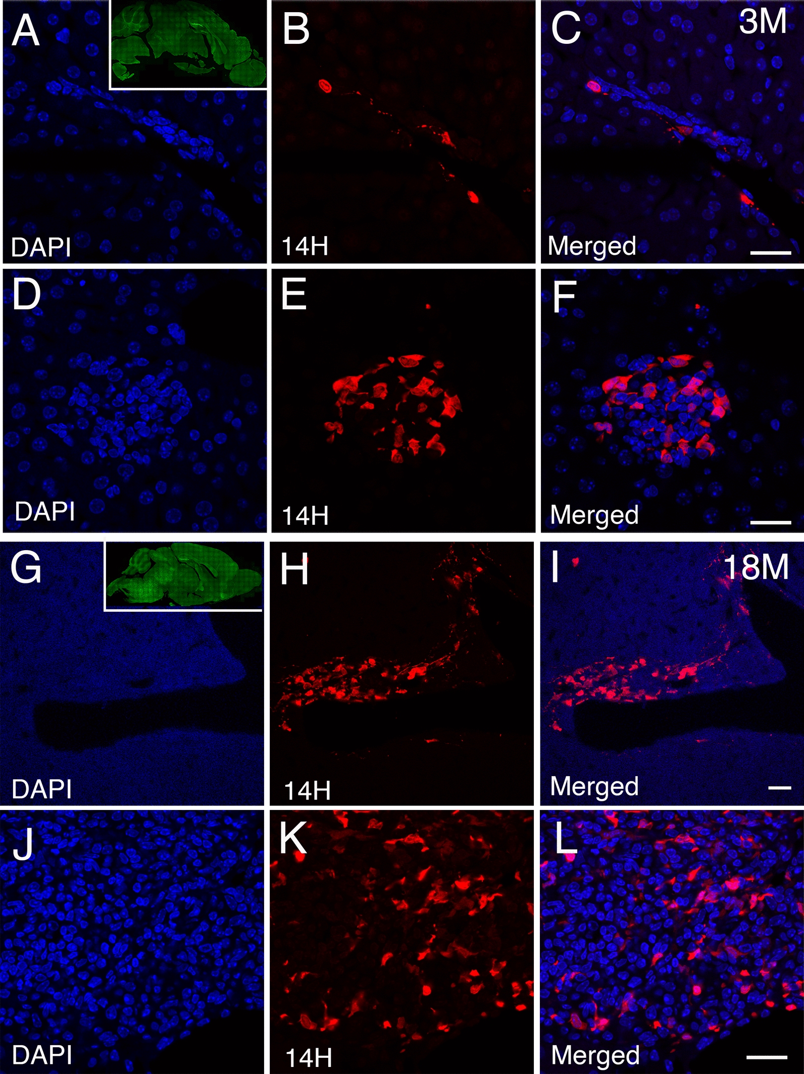
Age dependent accumulation of human α-syn deposits within the liver of the A30P transgenic mouse model. A–C Identification of human α-syn deposits (red) in young (3 months) liver tissue sections of the A30P mice located within the portal tracts and D–F, rosette-like structures within the liver parenchyma. Insert within panel A shows the deposition of α-syn at 3 months of age immune-stained with pS129 antibodies (green). G–I Aged A30P liver tissue sections (18 months) showing the progressive accumulation of α-syn deposits within the portal tracts and J–L, liver parenchyma. Insert within panel G shows the deposition of α-syn at 18 months of age immune-stained with pS129 antibodies (green). All liver tissue sections were counterstained with DAPI (blue). Bars = 20 µm