
Fig. 8.
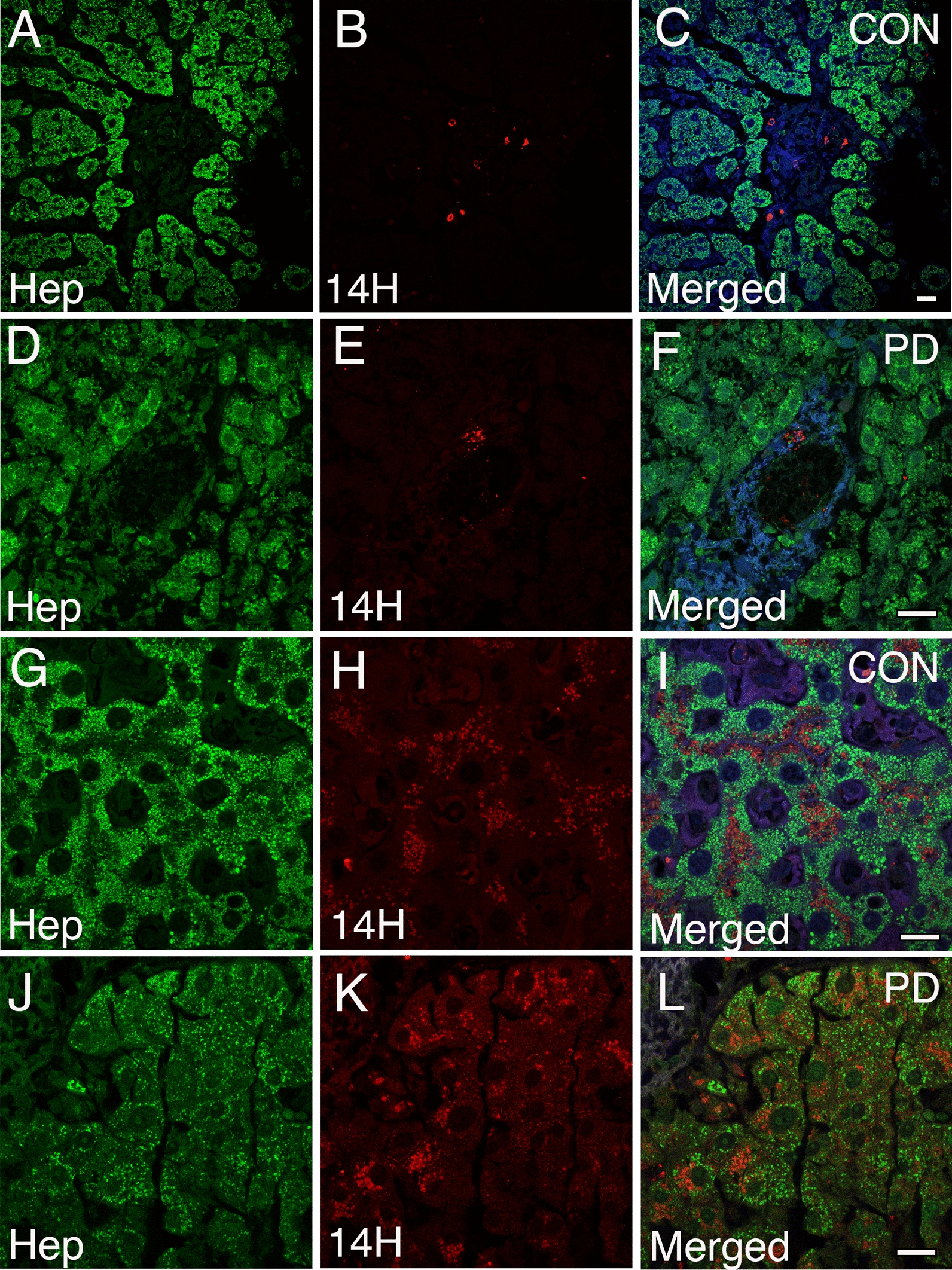
Accumulation of human α-syn within the human liver. A–C Confocal image analysis on human liver tissue sections immunolabeled with the 14H human-specific antibody (red) and anti-hepatocyte antibody (Hep, green) demonstrates the presence of human α-syn deposits within the sinusoidal regions, D–F portal tracts, and liver parenchyma (G–L) in PD and age-matched controls. Note that α-syn within liver hepatocytes appear as round puncta and often near nuclei. Importantly, not all hepatocytes within the affected region contain α-syn deposition. All tissue sections were counterstained with DAPI (blue). Bars = 20 µm