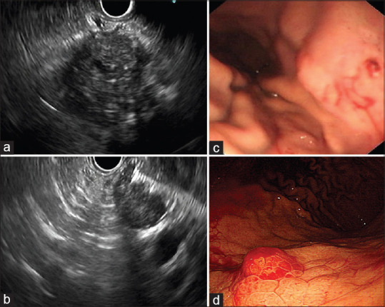
Figure 1.

EUS-FNA of the patient. (a) EUS showed diffuse enlargement of the pancreas, with hypoechoic parenchyma and multiple dot-like and linear hyperechoic lesions. (b) EUS-FNA was performed (once, ten strokes) with a 19-gauge needle. (c) No significant bleeding was observed just after the procedure. (d) An emergency upper endoscopy showed mucosal swelling at the upper posterior wall of the body of the stomach consistent with the puncture site, with bleeding on day 6 after EUS-FNA
