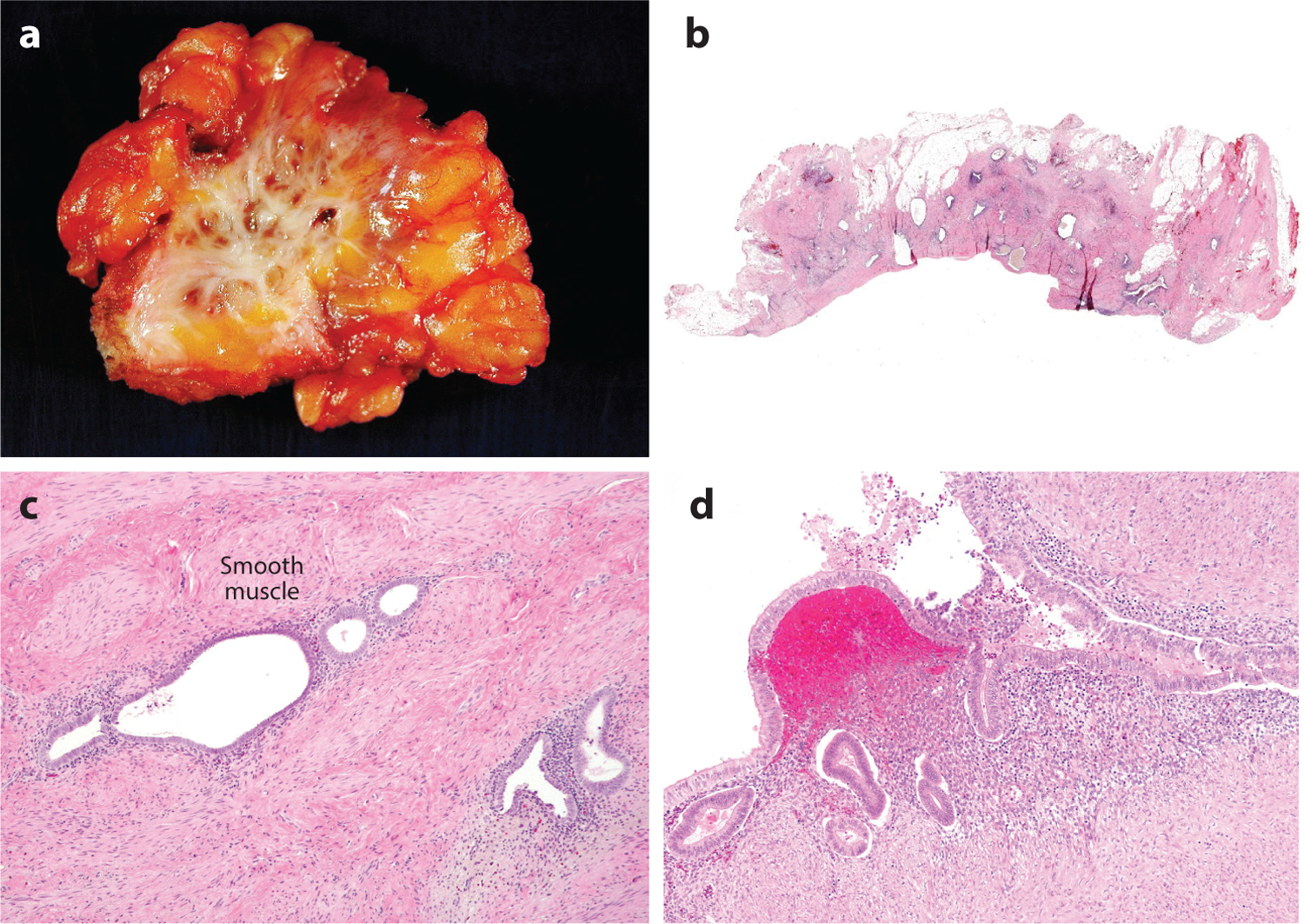
Figure 2.

Gross appearance and histology of deep infiltrating endometriosis (DIE). (a) DIE involves fibro-adipose tissue, resulting in extensive scarring. (b) Discrete endometrial glands surrounded by fibrosis are evident in sections from the DIE shown in panel a. (c) Hematoxylin and eosin–stained section of DIE shows the presence of glands and endometrial-type stroma in soft tissue. (d) An example of superficial endometriosis showing involvement of the surface of a fallopian tube.
