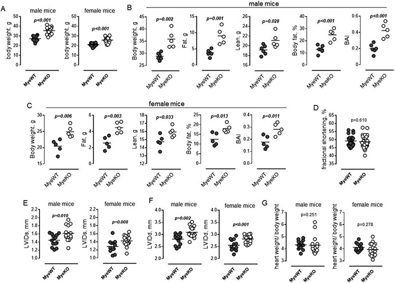
Figure 2. ErbB3MyeKO mice show higher weight but no differences in cardiac function at baseline.

A. Body weight of 15-week old male (left) ErbB3MyeWT (n=14) and ErbB3MyeKO (n=16) and female (right) ErbB3MyeWT (n=13) and ErbB3MyeKO (n=18) mice. Unpaired t test. B-C. Fat and lean tissue composition in male (B) and female (C) mice were determined using NMR as described in Methods. Body fat percentage was calculated by dividing fat mass by body weight; BAI (body adiposity index) as a ratio of fat mass to lean mass; n=5, unpaired t test. D. Fractional shortening in MyeWT (n=27) and MyeKO (n=34) animals (males and females). Unpaired t test. E-G. Left ventricular end-systolic (E) and end-diastolic diameters (F) in male (left) and female (right) mice. G. Heart-to-body weight ratios in male (left) and female (right) mice. E-G. n=14, male ErbB3MyeWT; n=16, male ErbB3MyeKO; n=13, female ErbB3MyeWT; n=18, female ErbB3MyeKO mice; D-H. Horizontal lines are indicating mean values. Statistical significance was calculated using unpaired t test.
