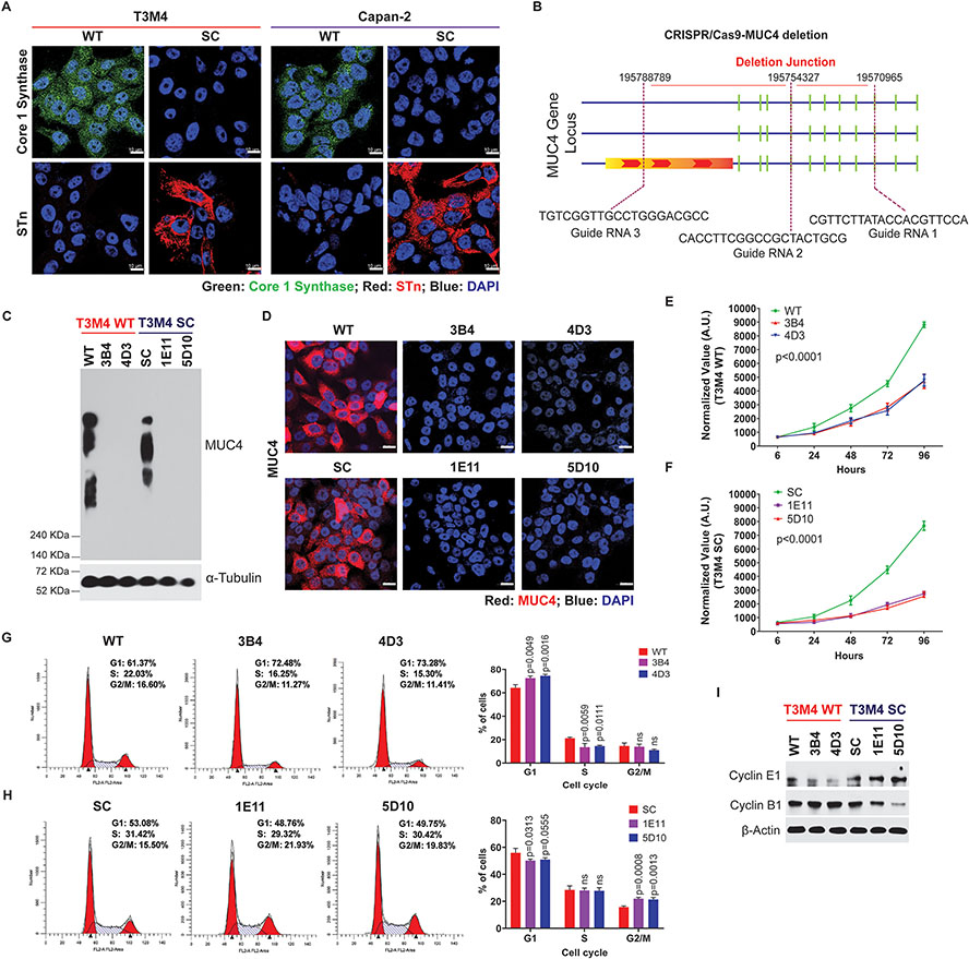
Figure 1. Genetic deletion of MUC4 reduces PDAC cell tumorigenicity.
A. Immunofluorescence analysis of core 1 synthase and STn in T3M4 and Capan-2 (WT and SC) cells. Scale bar (10μm). B. Schematic representation of MUC4 deletion using the CRISPR-Cas9 MUC4 gene construct. C. Immunoprobing of MUC4 in T3M4 WT, WT-MUC4KO (3B4 and 4D3), and T3M4 SC, SC-MUC4KO (1E11 and 5D10) cells. D. Immunofluorescence analysis of MUC4 in T3M4 WT, WT-MUC4KO (3B4 and 4D3), and T3M4 SC, SC-MUC4KO (1E11 and 5D10) cells. Scale bar (10μm). E and F Alamar blue cell proliferation assays for T3M4 WT, WT-MUC4KO (3B4 and 4D3), and T3M4 SC, SC-MUC4KO (1E11 and 5D10) cells at different time points (6, 24, 48, 72, and 96 h) (mean ± SD, n=3). G and H Cell cycle analysis of T3M4 WT, WT-MUC4KO (3B4 and 4D3), and T3M4 SC, SC-MUC4KO (1E11 and 5D10) cells by FACS (mean ± SD, n=3). I. Western blot analysis of Cyclin E1 and Cyclin B1 in T3M4 WT, WT-MUC4KO (3B4 and 4D3), SC, SC-MUC4KO (1E11 and 5D10) cells. β-actin acts as a loading control. [ns = not significant].

