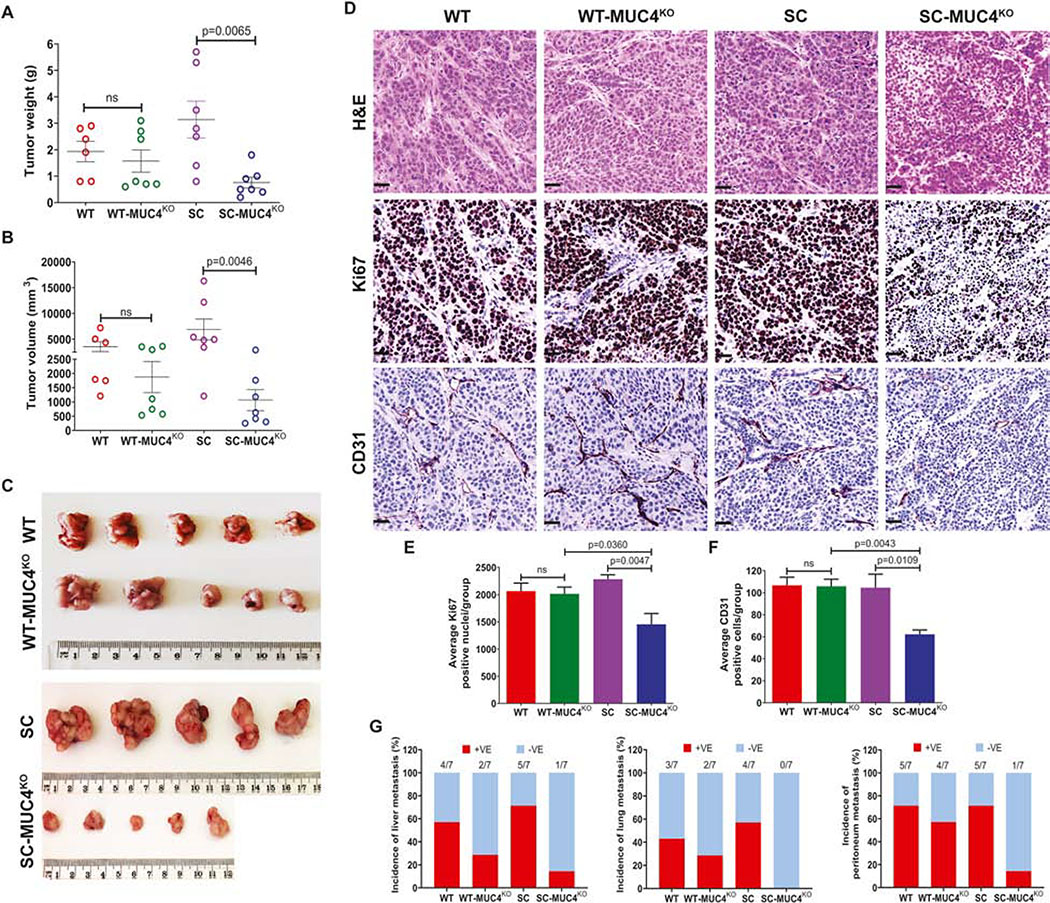
Figure 3. MUC4 deletion affects in vivo tumorigenesis.
A and B Quantification of tumor weight (g) and tumor volume (mm3) in T3M4 WT, WT-MUC4KO (4D3), SC, SC-MUC4KO (5D10) cells derived orthotopic tumors (n=7). C. Representative tumor images from the above-mentioned tumor models. D. Immunohistochemical analysis of H&E, Ki67 and CD31 in the above-mentioned tumor tissues. E. Average Ki67 positive cells (mean ± SD, n=5). F. Average CD31 positive cells (mean ± SD, n=5). G. Percentage of tumor metastasis (liver, lung, and peritoneum) in T3M4 WT, WT-MUC4KO (4D3), SC, SC-MUC4KO (5D10) cells implanted orthotopic tumor bearing-animals (n=7). [ns = not significant].

