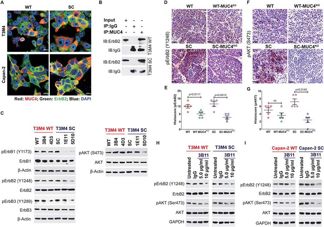
Figure 4. MUC4 mediates PDAC tumorigenicity through the regulation of ErbB2.
A. Co-localization of MUC4 (red) and ErbB2 (green) in T3M4 and Capan-2 (WT and SC) cells. Representative images are shown (n=3). Scale bar 10 μm. B. Immunoprecipitation (IP) of T3M4 (WT and SC) cell lysates with MUC4 (mAb 8G7) and mouse IgG (isotype control), and immunoprobed (IB) with anti-ErbB2 and anti-mouse IgG antibodies. C. Western blot analysis of phospho-ErbB1 (Y1173), ErbB1, phospho-ErbB2 (Y1248), ErbB2, phospho-ErbB3 (Y1289), and ErbB3, phospho-AKT (S473) and AKT in T3M4 WT, WT-MUC4KO, SC, and SC-MUC4KO cell lysate. β-actin acts as a loading control. IHC analysis of p-ErbB2 (D and E) and p-AKT (S473) (F and G) in T3M4 WT, WT-MUC4KO, SC, and SC-MUC4KO cells implanted orthotopic tumor tissues (n=5). H. Western blot analysis of p-ErbB2, ErbB2, p-AKT, and AKT in T3M4 WT and SC cells treated with varying doses of anti-Tn-MUC4 mAb 3B11 (5 and 10 μg/ml). I. Western blot analysis of p-ErbB2, ErbB2, p-AKT, and AKT in Capan-2 WT and SC cells treated with varying doses of anti-Tn-MUC4 mAb 3B11 (5 and 10 μg/ml) for 24 h. Treatment of PDAC cells with mouse IgG (5 μg/ml) served as an isotype control. GAPDH act as a loading control. [ns = not significant].

