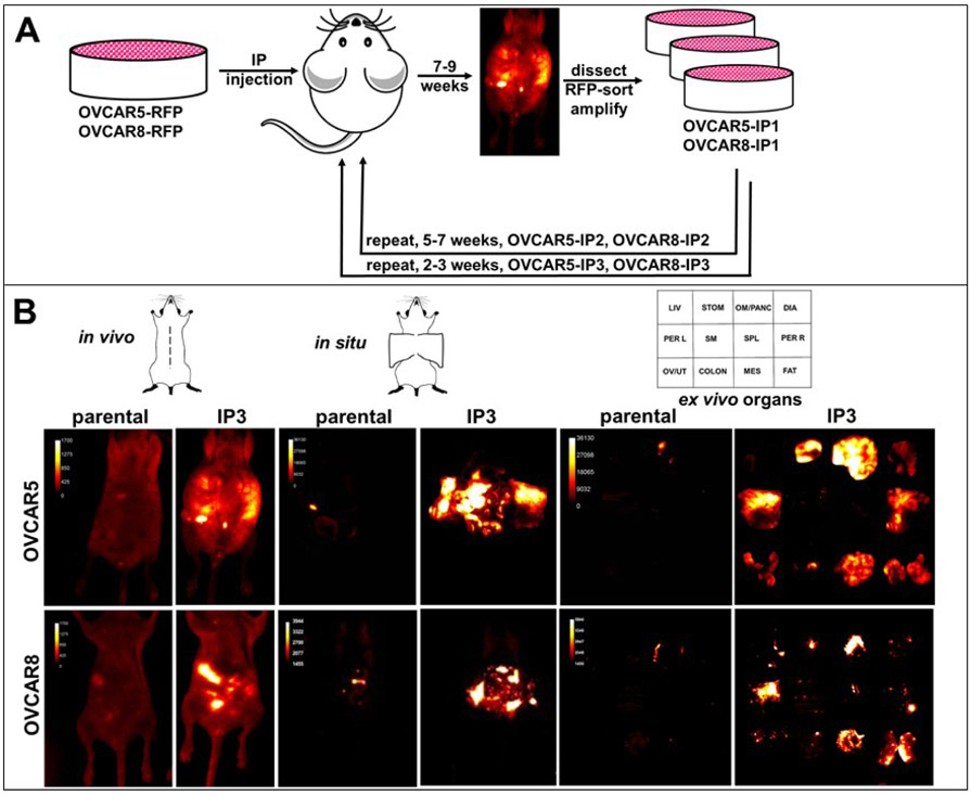
Figure 1. Serial in vivo passaging generates aggressive sublines of OVCAR5 and OVCAR8.
(A) Cells tagged with RFP (3x10⁶ in 1 ml PBS) were injected IP into female nu/nu mice and tumor burden monitored longitudinally by in vivo fluorescent imaging. When animals displayed significant tumor burden, they were sacrificed, tumor nodules dissected and cultured. Two additional rounds of in vivo selection were performed to generate the sublines OVCAR5-IP3 and OVCAR8-IP3 (n=4-6 per cohort). (B) Fluorescent images of tumors were obtained of the whole body (in vivo), midline dissected mice (in situ), or ex vivo organs (ex vivo). The template depicts the position of individual organs.

