
Fig. 1. ER stress mediates the killing effect of sulindac sulfide in HCT116 cells.
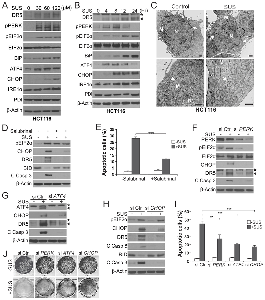
(A) Western blotting of indicated ER stress markers in HCT116 cells treated with sulindac sulfide (SUS) at indicated concentrations for 24 hr. (B) Western blotting of indicated ER stress markers in HCT116 cells treated with 120 μM SUS at indicated time points. *, non-specific bands. (C) HCT116 colon cancer cells treated with 120 μM SUS for 24 hr were analyzed by transmission electron microscopy (TEM). Arrows: endoplasmic reticulum; M: Mitochondria; N: nucleus. Scale bars: 1.0 µm. (D) Western blotting of indicated proteins in HCT116 cells treated with 120 μM SUS for 24 hr with or without pre-treatment with the ER stress inhibitor Salubrinal (1.0 μM) for 1.5 hr. C Casp 3, cleaved caspase 3. (E) Apoptosis in HCT116 cells treated with SUS for 48 hr with or without Salubrinal pre-treatment as in (D) was analyzed by counting condensed and fragmented nuclei after nuclear staining with Hoechst 33258. (F)-(J) HCT116 cells transfected with control scrambled, PERK, ATF4 or CHOP siRNA were treated with 120 μM SUS. (F)-(H) Western blotting of indicated proteins after SUS treatment for 24 hr. (I) Analysis of apoptosis after SUS treatment for 48 hr as in (E). (J) Crystal violet staining of viable cells after SUS treatment for 48 hr. Results in (E) and (I) were expressed as means + SD of three independent experiments. **P < 0.01; ***P < 0.001.