
Fig. 3. ER stress mediates the killing effect of sulindac sulfide on non-transformed colonic epithelial cells with APC loss.
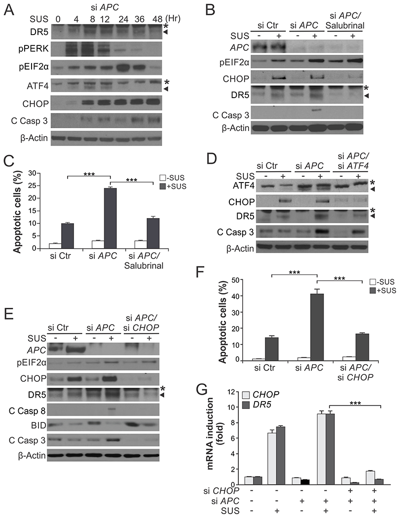
(A) Western blotting of indicated proteins in NCM356 normal colonic epithelial cells transfected with APC siRNA and then treated with 120 μM sulindac sulfide (SUS) at indicated time points. C Casp 3, cleaved caspase 3. *, non-specific bands. (B) Western blotting of indicated proteins in NCM356 cells transfected with control scrambled or APC siRNA with or without treatment with 120 μM SUS for 48 hr and pre-treatment with the ER stress inhibitor salubrinal (1.0 μM) for 1.5 hr. *, non-specific bands. (C) Apoptosis in NCM356 cells treated as in (B) was analyzed by counting condensed and fragmented nuclei after nuclear staining with Hoechst 33258. (D)-(G) NCM356 cells were transfected with indicated siRNA or combinations, and then treated with 120 μM SUS for 48 hr. (D), (E) Western blotting of indicated proteins. (F) Analysis of apoptosis as in (C). (G) RT-PCR analysis of mRNA expression of indicated genes. Results in (C), (F) and (G) were expressed as means + SD of three independent experiments. ***P < 0.001.