
Fig. 5. Inhibition of ER stress suppresses the chemopreventive and apoptotic effects of sulindac in APCMin/+ mice.
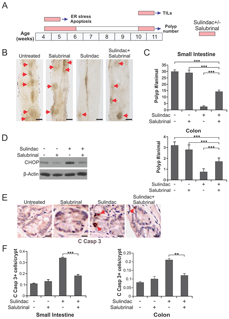
(A) Schemes of sulindac (dietary; 200 ppm) and salubrinal (IP; 1 mg/kg/day) treatments for analyzing ER stress and apoptosis, polyp numbers, and TILs in APCMin/+mice. (B), (C) Four-week-old APCMin/+mice were treated with sulindac +/− salubrinal as in (A) for analyzing polyp numbers. (B) Representative images of the small intestine of control and treated mice, with arrows indicating microscopic lesions (polyps/adenomas). Scale bars: 2 mm, n=4. (C) Mean numbers + SD of adenomas (>0.5mm in diameter) in the small intestine (upper panel) and colon (lower panel). (D)-(F) Four-week-old APCMin/+mice treated with sulindac +/− salubrinal as in (A) for analyzing ER stress and apoptosis. (D) Western blotting of CHOP in the small intestinal mucosa isolated from the treated mice. (E) Representative pictures of active caspase 3 (C Casp 3) immunostaining of small intestinal sections, with arrows indicating example cells with positive staining. Scale bars: 25 µm. (F) Quantification of active caspase signals in the sections of small intestine (left panel) and colon of the treated mice (right panel). In (C) and (F), means + SD are shown, n=3. **P <0.01; ***P < 0.001