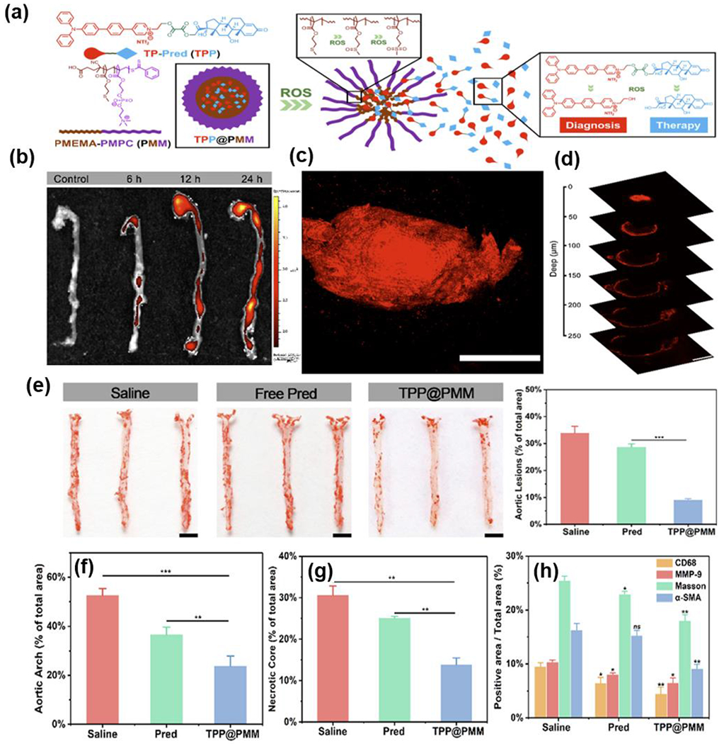
Fig.10.

(a) Illustration of developing a nanoplatform with two-photon imaging. (b) Ex vivo fluorescence images and quantitative result of TPP@PMM accumulation in aortas. (c) Two-photon confocal image of the atherosclerotic plaques. (d) Two-photon CLSM images of the plaques at various imaging depths. (e) Photographs of en face Oil red O-stained aortas and quantitative result of the Oil red O positive areas from the mice treated with different formulations. (f) Quantitative analysis of the plaque area, (g) necrotic core area and (h) positive area in different histochemistry analyses. Reproduced with permission from Ref. [337]. Copyright 2020, American Chemistry Society.
