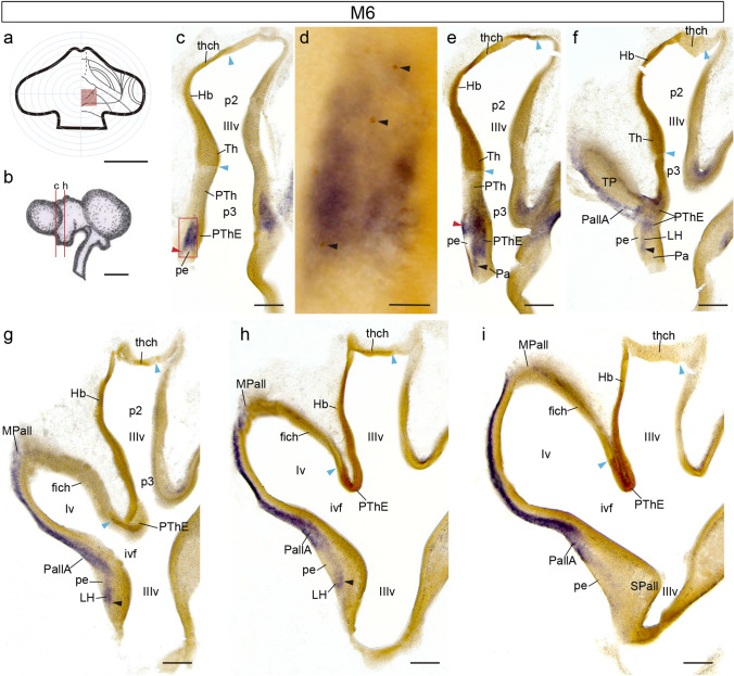
Fig. 2.
Coronally sectioned chimeric embryo (case M6), fixed at stage HH25/26, and processed for Tbr1 ISH and QCPN IMR. a Drawing of graft extent relative to the prosencephalic fate map. b Schema of the M6 chimeric brain, illustrating the plane of the sections shown in (c, e–i). c, e–i Caudorostral series of coronal sections through the di-telencephalic transition. Blue arrowheads indicate the borders of the graft at the ventricle, red arrowheads point to the peripeduncular stream, and black arrowheads point to the juxtapeduncular stream. d Magnification of the area framed in (c). Black arrowheads point to quail-derived cells in the PThE. Scale bars in a 250 μm, b, 2.5 mm, c, e-i, 150 μm, and d, 50 μm

