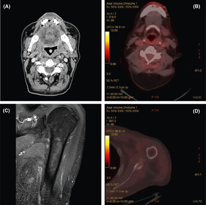
FIGURE 2.

A: Computed tomography (CT) scan (laryngeal windows) of the patient after administration of intravenous contrast media. Axial CT image showing an almost complete resolution of the solid lesion and complete lymph nodal response. B: PET‐CT showing complete response after therapy in head and neck district. C: Magnetic resonance after therapy showing complete resolution of the lesion. D: PET‐CT showing complete response after therapy in the humerus
